You are here: Urology Textbook > Surgical Management > Ureteral stent
Ureteral Stents (DJ and MJ): Indications, Technique, and Complications
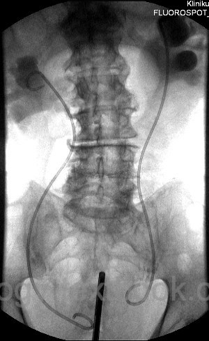
Ureteral stents are thin catheters placed within the ureter to ensure urinary drainage from the renal pelvis. They maintain urinary transport along the ureter and bypass obstructions. Internal ureteral stents drain urine from the renal pelvis into the bladder [Fig. DJ stent], whereas external ureteral stents divert the urine into a collection bag via the urethra or transcutaneously.
Synonyms For Ureteral Stents:
Ureteral catheter, ureteral splint, ureteral stent, double-J or DJ stent.
 |
Internal Ureteral Stents
Definition:
An internal ureteral stent is a thin, flexible tube in the ureter with two curled ends located in the renal pelvis and bladder to maintain its position. Due to its characteristic shape, it is also known as a double-J or DJ stent.
Indications for internal ureteral stenting:
Indications are summarized below. Long-term placement of ureteral stents requires regular exchange every 3–6 months, or earlier in cases of incrustation or recurrent obstruction.
- Acute obstruction: ureteral stones, upper urinary tract bleeding with clot formation, ureteral compression from hematoma or abscess.
- Urinoma: caused by fornix rupture (e.g., due to ureteral stone), iatrogenic ureteral trauma (URS, surgery), or renal trauma with urinary extravasation.
- Chronic hydronephrosis: advanced tumors causing ureteral compression, retroperitoneal fibrosis (Ormond disease), and ureteral strictures.
- Prophylactic stenting: before ESWL of large stones, before ureteroscopy to dilate the ureter, after URS, before renal surgery (partial nephrectomy, pyeloplasty), or before abdominal tumor resections with ureteral displacement.
Contraindications for Internal Ureteral Stenting
- Inability to place the patient in the lithotomy position
- Loss of bladder function, e.g., by tumor infiltration
- In cases of technical difficulty, repeated attempts should be weighed against the need for immediate percutaneous nephrostomy, especially in the context of urosepsis.
Design and Materials of Internal Ureteral Stents
Stents are commonly made of polyurethane, silicone, or proprietary polymers (C-flex, Silitek, Percuflex, Tecoflex). The material determines flexibility (memory effect), surface properties, and the tendency for incrustation. Randomized comparative trials between different materials are unavailable. Stents differ in shape, length, diameter, and technical features:
Shape:
Both ends curl (“J-shaped”) to anchor the stent in the renal pelvis and bladder. Side holes allow urine drainage. Some models are open-ended, enabling wire-guided exchanges, while others are closed-ended.
Length:
The length of the ureteral stent is jugded with the retrograde pyelography. In adults, the typical length ranges from 26 to 34 cm, depending on the distance between the bladder and renal pelvis and ureteral tortuosity. The measuring reference points may vary between different manufacturers.
Diameter:
Measured in French (Fr) or Charrière (Ch), the most commonly used ureteral stents are 6–8 Fr in diameter. Thin stents are used prophylactically, for example, after ureteroscopic treatment of ureteral stones (nephrolithiasis). Thicker stents are indicated in cases of upper urinary tract bleeding, after treatment of ureteral strictures, or in patients with rapid incrustation of ureteral stents.
Special Features:
In addition to the above-mentioned basic properties of ureteral stents, numerous other technical variations are helpful in specific situations:
- Extraction string at the distal end
- Pushers and guiding systems for placement
- Hydrophilic coating to ease insertion
- Anti-reflux valves
- Metal-reinforced spirals against tumor compression
Technique of DJ Stent Placement
After retrograde pyelography, a guidewire is advanced into the renal pelvis using the working channel of the cystoscope. The stent is then advanced over the wire using a pusher until the proximal curl is placed into the renal pelvis. Withdrawal of the guide wire allows the distal end to coil in the bladder. In difficult cases (e.g., strictures, kinking), hydrophilic or stiff wires may be necessary. If retrograde stenting fails, percutaneous nephrostomy or antegrade insertion of the ureteral stent are the remaining options.
Complications of internal stents
The most frequent complications are irritative voiding symptoms (frequency, urgency, dysuria) and flank pain due to vesicoureteral reflux.
Incrustation:
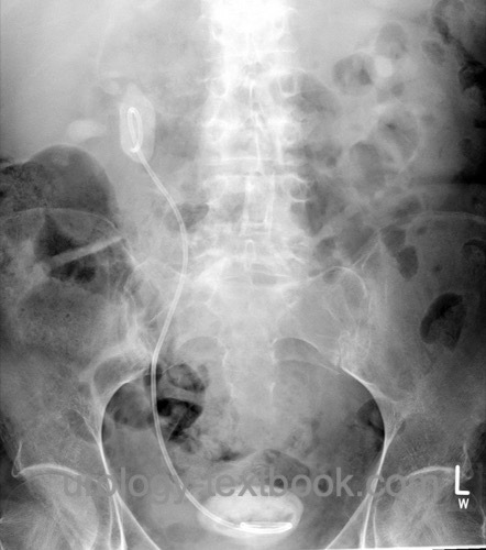
Biofilm leads to encrustation and obstruction. Forgotten stents may be encased in massive stone formations, making removal difficult [Fig. Forgotten DJ stent].
 |
Dislocation:
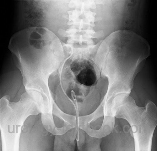
Cranial migration (distal end slips into the ureter) causes obstruction. Kaudal migration leads to incontinence if the distal end passes the external urinary sphincters [fig. dislocated stent].
 |
Other complications:
Infections, urinomas, and rarely fistulization into adjacent structures (bowel, vessels).
Removal of Internal Ureteral Stents
- Removal is typically performed using a grasping forceps through the working channel of the cystoscope under local anesthesia. Stent removal may be combined with retrograde pyelography or second-look URS in general anesthesia.
- Easy stent removal after short-term stenting is possible with the help of the extraction string: the string exits the urethra after the procedure and is secured to the patient's thigh or abdomen for a few days ("stent on a string").
External Ureteral Stents
Diagnostic Ureteral Catheters:
Ureteral Catheters (3–10 Fr) are used for ureteral probing and retrograde pyelography. Open catheter tips (straight or curved) facilitate the easy insertion of a guide wire for coaxial working techniques following retrograde pyelography. Other tip shapes include the olive tip (facilitates probing) and the Chevassu tip (conical thickening) for optimized retrograde pyelography.
MJ Ureteral Stents for Urinary Drainage:
An MJ ureteral stent is a thin, flexible tube in the ureter with one curled end located in the renal pelvis. The distal straight end is externalized via the urethra, percutaneously, or a stoma, and drains into a urine bag. Transurethral MJ stents are sutured to a thin Foley catheter for fixation. They are easy to remove postoperatively and allow flushing in cases of clot or pus obstruction. Since transurethral MJ stents are uncomfortable and prone to dislocation, they are only indicated for a short time in the following situations:
- Short-term drainage after ureteroscopy
- Drainage of severe pyuria or hematuria with clot obstruction
- After surgery of the ureter, e.g., ureteral reimplantation.
- Chronic or permanent use: stomal stenosis of ureterocutaneostomy, ureteroileal anastomotic stenosis in ileal conduit diversion
Ureteral occlusion catheters:
A ureteral occlusion catheter is a single- or double-lumen polyurethane catheter with balloon occlusion. It is used to block the ureter before percutaneous nephrolithotomy to prevent stone migration and to facilitate puncture.
Complications of external stents:
- Accidental dislodgement due to traction
- Ascending urinary tract infections
- Urethral pain, urethral infection
- Other complications are similar to those of internal stents, particularly when used for an extended period.
| Intermittent catheter | Index | Percutaneous nephrostomy |
Index: 1–9 A B C D E F G H I J K L M N O P Q R S T U V W X Y Z
References
Ahmad MU, Siddiqui S, Ashraf FA, Iqbal R, Ehsanullah SAM, AlFayadh A, Siddiqui MRS, Khan MS, Furrer MA. Retrograde Ureteral Stents Versus Percutaneous Nephrostomy in the Management of Malignant Ureteral Obstruction: A Systematic Review and Meta-Analysis. Urology. 2024 Oct;192:158-167. doi: 10.1016/j.urology.2024.05.042.
Cardoso A, Coutinho A, Neto G, Anacleto S, Tinoco CL, Morais N, Cerqueira-Alves M, Lima E, Mota P. Percutaneous nephrostomy versus ureteral stent in hydronephrosis secondary to obstructive urolithiasis: A systematic review and meta-analysis. Asian J Urol. 2024 Apr;11(2):261-270. doi: 10.1016/j.ajur.2023.03.007.
Fiuk J, Bao Y, Calleary JG, Schwartz BF, Denstedt JD. The use of internal stents in chronic ureteral obstruction. J Urol. 2015 Apr;193(4):1092-100. doi: 10.1016/j.juro.2014.10.123.
Tomer N, Garden E, Small A, Palese M. Ureteral Stent Encrustation: Epidemiology, Pathophysiology, Management and Current Technology. J Urol. 2021 Jan;205(1):68-77. doi: 10.1097/JU.0000000000001343.
Wang X, Ji Z, Yang P, Li J, Tian Y. Forgotten ureteral stents: a systematic review of literature. BMC Urol. 2024 Mar 5;24(1):52. doi: 10.1186/s12894-024-01440-9.
 Deutsche Version: Indikationen und Komplikationen von Harnleiterschienen (DJ und MJ Stents)
Urology-Textbook.com – Choose the Ad-Free, Professional Resource
This website is designed for physicians and medical professionals. It presents diseases of the genital organs through detailed text and images. Some content may not be suitable for children or sensitive readers. Many illustrations are available exclusively to Steady members. Are you a physician and interested in supporting this project? Join Steady to unlock full access to all images and enjoy an ad-free experience. Try it free for 7 days—no obligation.
New release: The first edition of the Urology Textbook as an e-book—ideal for offline reading and quick reference. With over 1300 pages and hundreds of illustrations, it’s the perfect companion for residents and medical students. After your 7-day trial has ended, you will receive a download link for your exclusive e-book.