You are here: Urology Textbook > Kidneys > Renal abscess
Renal Abscess: Symptoms, Diagnosis and Treatment
Review literature: (Angel et al., 2003) (Shu et al., 2004).
Definition of a Renal Abscess
A renal abscess is a collection of pus within the kidney. The kidney infection may extend into the retroperitoneum.
Etiology of Renal Abscess
Gram-negative Bacteria:
A renal abscess is most commonly caused by gram-negative bacteria such as E. coli and Proteus. Ascending urinary tract infections (pyelonephritis), tubular obstruction, and nephrolithiasis are common risk factors for renal abscess.
Staphylococci:
Before the antibiotic era, renal abscesses were often caused by the hematogenous spread of staphylococci. Today's risk groups for the hematogenous spread of staphylococci are intravenous drug abuse, intensive care, dialysis patients, valvular heart disease, and endocarditis.
.Signs and Symptoms of a Renal Abscess
- Fever and chills
- Flank pain or abdominal pain
- Weight loss, malaise
- Urosepsis
| Do you want to see the illustration? Please support this website with a Steady membership. In return, you will get access to all images and eliminate the advertisements. Please note: some medical illustrations in urology can be disturbing, shocking, or disgusting for non-specialists. Click here for more information. |
Kidney Abscess: Diagnostic Workup
Urine culture:
Urine culture can often isolate the responsible bacteria and enables antibiotic resistance testing.
Laboratory tests:
Blood culture (positive in 50%). Blood count, coagulation tests (PTT, PT), CRP, liver enzymes, creatinine. In suspected urosepsis, determine sepsis parameters such as blood gas testing, procalcitonin, AT III and fibrinogen.
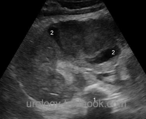
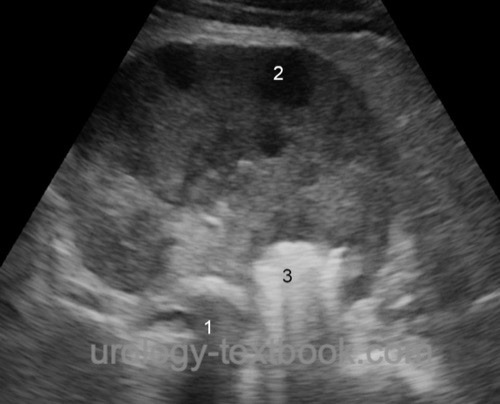
Ultrasonography:
Sonographic signs for a renal abscess in renal ultrasound are a hypoechoic mass within the renal capsule, which may have inclusions of air (echogenic reflex with dorsal shadowing). Doppler sonography may show increased blood flow at the margin of the renal abscess, whereas blood flow is often not visible in the center of the mass. CT should be initiated in case of abnormalities in the sonography.
 |
 |
 |
Computed tomography:
A CT scan is the diagnostic test of choice [fig. renal abscess]. The renal abscess appears as a hypodense area. Air inclusion is possible. After administration of contrast medium, the abscess capsule has a ring-like enhancement.
 |
Intravenous urography:
Intravenous urography has only historical value and is now replaced by CT. Urography may be normal and may fail to diagnose renal abscess. If done, KUB X-ray may show an increased renal shadow and nephrolithiasis. The excretion of contrast medium is often delayed with a displacement of the pyelocalyceal system.
Treatment of a Renal Abscess
Cornerstones in the treatment of renal abscess are parenteral antibiotics and abscess drainage:
- Parenteral antibiotics: e.g., amoxicillin and clavulanic acid 2,2 g 1–1–1 IV in combination with gentamicin 3 mg/kg 1–0–0 IV Alternatives are third-generation cephalosporins.
- Abscess drainage: renal abscess larger than 3 cm diameter should be drained. Pus is sent for identification of bacteria and antibiotic resistance testing. Renal abscess larger than 5 cm diameter may need repeated or several parallel pigtail drains.
- In advanced disease, surgical drainage or nephrectomy is rarely necessary, depending on renal function.
| Chronic pyelonephritis | Index | Perinephric abscess |
Index: 1–9 A B C D E F G H I J K L M N O P Q R S T U V W X Y Z
References
Angel u.a. 2003 ANGEL, C. ; SHU, T. ; GREEN, J. ; ORIHUELA, E. ; RODRIQUEZ, G. ; HENDRICK, E.: Renal and peri-renal abscesses in children: proposed physio-pathologic mechanisms and treatment algorithm.In: Pediatr Surg Int
19 (2003), Nr. 1–2, S. 35–9
Shu u.a. 2004 SHU, T. ; GREEN, J. M. ;
ORIHUELA, E.:
Renal and perirenal abscesses in patients with otherwise anatomically
normal urinary tracts.
In: J Urol
172 (2004), Nr. 1, S. 148–50
 Deutsche Version: Nierenabszess
Urology-Textbook.com – Choose the Ad-Free, Professional Resource
This website is designed for physicians and medical professionals. It presents diseases of the genital organs through detailed text and images. Some content may not be suitable for children or sensitive readers. Many illustrations are available exclusively to Steady members. Are you a physician and interested in supporting this project? Join Steady to unlock full access to all images and enjoy an ad-free experience. Try it free for 7 days—no obligation.
New release: The first edition of the Urology Textbook as an e-book—ideal for offline reading and quick reference. With over 1300 pages and hundreds of illustrations, it’s the perfect companion for residents and medical students. After your 7-day trial has ended, you will receive a download link for your exclusive e-book.