You are here: Urology Textbook > Examinations > Abdominal x-ray
Indications and Normal Findings of Abdominal X-Ray
Indications for Abdominal X-Ray
- Before IV urography (plain film)
- Abdominal pain or acute abdomen,
- Gastrointestinal obstruction, suspected bowel perforation
- Nephrolithiasis
- Search for foreign bodies
Abdominal X-ray has clear disadvantages in sensitivity and specificity compared to an abdominal CT in almost all issues.
 |
Examination Technique
For urological questions, imaging is done in anteroposterior (AP) projection, which leads to a better representation of the retroperitoneal structures (KUB = X-ray of kidneys, ureters, and bladder). In the case of an acute abdomen or gastrointestinal obstruction, the posterior-anterior projection is chosen for better imaging of the intraperitoneal organs.
Coverage on the X-ray should include the 11th thoracic vertebrae and diaphragm cranially. Caudally, the lower margin of the symphysis should be visible. Two images may be necessary for large patients. Depending on the question and prior findings, the target area of the image can be reduced (for example, half-sided KUB after stone therapy).
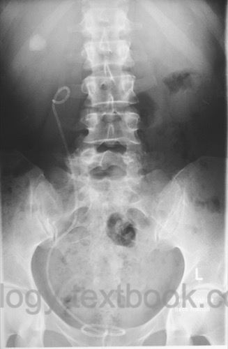
Normal Findings
In good examination conditions, the kidneys are visible. The length is usually 11–12 cm, and the width is around 5 cm. The upper pole of the left kidney is at the upper edge of the 12th thoracic vertebra, the lower pole ends at the middle of 3rd lumbar vertebra. The right kidney is shifted about 1.5 cm to the pelvis.
Deviations from the typical renal shape may be caused by renal cysts, renal infarctions, kidney tumors, abscess, or congenital malformations. Calcifications in projection on the urinary tract are not only caused by urinary calculi; consider also lymph node calcifications, phleboliths, arteriosclerosis, chronic pancreatitis, intestinal contents, or foreign bodies.
The spine, ribs and pelvis should be carefully evaluated (fractures? cortex interruptions? ossal lesions?). Urological tumors often cause skeletal metastases. The psoas shadow should be symmetrical, a lost or asymmetric psoas shadow may be caused by retroperitoneal bleeding, tumor or aortic aneurysm. Assess the intestinal gas distribution for signs of obstruction.
| Urologic Surgery | Index | Intravenous Urography |
Index: 1–9 A B C D E F G H I J K L M N O P Q R S T U V W X Y Z
References
 Deutsche Version: Röntgen des Abdomens
Urology-Textbook.com – Choose the Ad-Free, Professional Resource
This website is designed for physicians and medical professionals. It presents diseases of the genital organs through detailed text and images. Some content may not be suitable for children or sensitive readers. Many illustrations are available exclusively to Steady members. Are you a physician and interested in supporting this project? Join Steady to unlock full access to all images and enjoy an ad-free experience. Try it free for 7 days—no obligation.
New release: The first edition of the Urology Textbook as an e-book—ideal for offline reading and quick reference. With over 1300 pages and hundreds of illustrations, it’s the perfect companion for residents and medical students. After your 7-day trial has ended, you will receive a download link for your exclusive e-book.