You are here: Urology Textbook > Examinations > Intravenous urography
Intravenous Urography: Technique and Normal Findings
Abbreviations and synonyms: IVP (intravenous pyelography), IVU (intravenous urography).
Indications for Intravenous Urography
The indications for intravenous urography are being pushed back by computed tomography (native or with contrast medium), which is superior in terms of sensitivity and specificity in almost every question. Without the availability of computed tomography, urography is a diagnostic option in the following situations: nephrolithiasis, gross hematuria, recurrent urinary tract infections, nephrolithiasis, urinary retention, urothelial carcinoma of bladder, upper urinary tract carcinoma, renal trauma, and urinary tract malformations.
 |
Examination Technique of Intravenous Urography
Premedication:
Premedication may reduce the rate of side effects of the iodinated contrast medium in certain situations: chronic kidney disease, allergic predisposition, or hyperthyroidism, see section pharmacology of iodinated contrast medium. Bowel preparation before IVP delays the examination and fails to show any clear benefit in randomized trials.
Examination technique:
After emptying the bladder, the examination begins with an abdominal X-ray (KUB, plain film). contrast medium is given if good examination conditions are seen (no meteorism or contrast medium remnants of preliminary studies).
1–1.5 ml/kg contrast medium with a concentration of 300 mg/ml of iodine is given with a rapid infusion. After bolus injection, further KUB radiographs are done after 3 min (collimated to the kidneys and best done with nephrotomography), after 6 min, and after 12 min [fig. intravenous urography with distal ureteral stone]. Improved imaging of the upper urinary tract is possible with an abdominal compression of the ureters after the 6 min KUB if no obstruction is present.
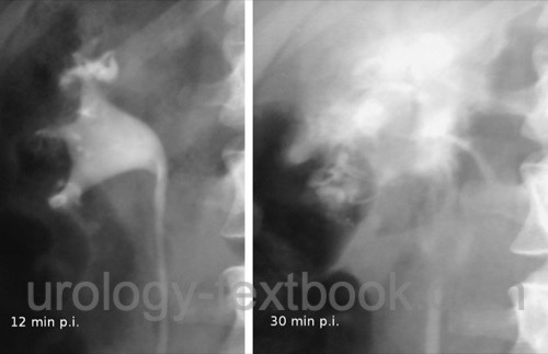
Depending on the results of the 6 and 12 min imaging, further radiographs are done to e.g., evaluate hydronephrosis with insufficient contrast [fig. IV urography of third-grade hydronephrosis]. Collimation should be adapted to the clinical question (lower abdomen, half-sided KUB). Additional radiographs to image bladder tumors or residual urine are no longer performed due to the wide availability of ultrasound imaging.
 |  |
Normal Findings of Intravenous Urography
Plain film KUB:
See section abdominal X-ray for normal findings.2--3 min KUB:
The nephrographic effect should be symmetric, without parenchymal defects, and the lateral convex border of the kidney is smooth. The renal length is usually 11–12 cm, and the width is around 5 cm. Parenchyma is 2–3 cm thick.
6 min KUB:
Usually the renal pelvis system and the proximal ureter are contrasted on both sides without filling defects or signs of obstruction. The anatomy of the calyces and papillae is described in fig. radiological anatomy of the renal calyces. See also section anatomy of the ureter and renal pelvis for the normal anatomy of the renal pelvis with its dendritic or ampullary norm variants.
 |
12 min KUB:
Normal findings are the complete contrasting of the ureters and the bladder without filling defects. Segmental narrowing of the ureter is caused by peristalsis. The diameter of the ureter proximal to the narrowing should be less than 8 mm. Signs of urinary obstruction are delayed contrast medium excretion, rounded calyceal fornices, calyceal "clubbing" and dilatation [fig. radiological signs of hydronephrosis], enlarged renal pelvis, and a ureter diameter >8 mm.
 |
Later KUB:
Later images are necessary if urinary obstruction causes delayed contrasting of the urinary tract. KUB after 1–2 hours help to find the level of obstruction and to judge the clinical significance of obstruction.
Complications of Intravenous Urography
Forniceal rupture:
Contrast medium increases diuresis and may cause a forniceal rupture [fig. forniceal rupture], most common in patients with an ureteral calculi. Forniceal rupture necessitates ureteral stenting. Intravenous urography should not be done in patients with renal colic.
 |
Complications due to contrast medium:
See section contrast medium.
| Abdominal X-ray | Index | VCUG |
Index: 1–9 A B C D E F G H I J K L M N O P Q R S T U V W X Y Z
References
 Deutsche Version: Intravenöse Urographie
Urology-Textbook.com – Choose the Ad-Free, Professional Resource
This website is designed for physicians and medical professionals. It presents diseases of the genital organs through detailed text and images. Some content may not be suitable for children or sensitive readers. Many illustrations are available exclusively to Steady members. Are you a physician and interested in supporting this project? Join Steady to unlock full access to all images and enjoy an ad-free experience. Try it free for 7 days—no obligation.
New release: The first edition of the Urology Textbook as an e-book—ideal for offline reading and quick reference. With over 1300 pages and hundreds of illustrations, it’s the perfect companion for residents and medical students. After your 7-day trial has ended, you will receive a download link for your exclusive e-book.