You are here: Urology Textbook > Urologic examinations > Imaging > Renal ultrasound
Renal Ultrasound: Sonography of the Kidneys
Ultrasonography of the kidneys is frequently indicated in urological patients. Indications include (but are not limited to): determination of size and localization of the kidneys, abdominal pain, flank pain, hematuria, proteinuria, lower urinary tract symptoms, abdominal trauma, (recurrent or febrile) urinary tract infections, increased retention parameters, nephrolithiasis, diagnosis and follow-up of renal tumors and cysts, kidney transplantation, planning and performing surgical procedures such as renal biopsy or percutaneous nephrolithotomy. Review literature: (Singer et al., 2006).
Examination Technique
The transducer used for renal ultrasonography is a sector or curved array probe with 3.5–5 MHz. The examination of the kidneys is done in supine position and with inspiration of the patient. In case of unfavorable conditions, a lateral decubitus position is helpful, whereby the side to be examined is raised by 30 degrees. Each kidney is screened in sagital and transverse plane [fig. normal findings in renal ultrasonography]. The upper pole is further dorsal than the lower pole, the transducer must be tilted accordingly.
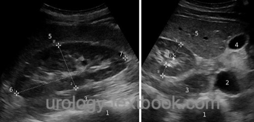 |
Normal Findings in Renal Ultrasound
Size if the Kidney
- Length: 100–115 mm
- Width: 50–70 mm
- Thickness: 30–50 mm
The size of the parenchyma is measured from the convex outer edge to the tip of a papilla, the normal value is 13–18 mm. The kidney volume of 110–200 ml and can be calculated by using the following formula:
| kidney volume | = | length × width × depth × 0.5 |
Renal parenchyma pyelon index:
The relationship between the dorsal parenchyma to pyelon to ventral parenchyma is normally 1:1:1. With increasing age, the kidney atrophy shifts the renal parenchyma pyelon index to 1:2:1.
Renal Ultrasound Anatomy
The kidney is a bean-shaped organ, which has usually a smooth convex lateral organ boundary. The renal parenchyma is homogeneously hypoechoic, sometimes the medullary pyramids are somewhat less echogenic than the cortex. The surrounding fat tissue and the pyelocalyceal system with parapelvic fat is hyperechoic. Medial to the pyelocalyceal system, sections of the great vessels and the psoas muscle are visible.
Contrast enhanced ultrasound (CEUS):
CEUS is indicated for the evaluation of cystic lesions of the kidney and for suspected renal infarction.
Doppler-Sonography of the Renal Vessels
Indications for renal Doppler sonography:
- Suspected renal artery stenosis or renal hypertension
- Suspected renal infarction
- Assessment of renal function after transplantation
Standard flow rates of the renal artery:
- Peak systolic velocity (PSV): 80–150 cm/s
- End-diastolic velocity (EDV): 20–50 cm/s
Resistive index of the kidney
The resistive index (RI) is calculated with the peak systolic velocity (PSV or VPSV) and the end-diastolic velocity (EDV or VEDV) using following formula:
RI=(VPSV-VEDV)/VPSV
The location for the assessment of PSV and EDV are the arcuate arteries or interlobar arteries at the border between cortex and medulla. The standard value for the resistive index is 0.5 to 0.7. A RI of >0.7 or a side difference of more than 0.1 are seen in hydronephrosis, renal transplant graft rejection or intrinsic renal disease. An RI of less than 0.5 is a sign for a renal artery stenosis.
| Doppler ultrasound | Index | Ureter ultrasound |
Index: 1–9 A B C D E F G H I J K L M N O P Q R S T U V W X Y Z
References
Singer u.a. 2006 SINGER, Eric A. ; GOLIJANIN, Dragan J. ; DAVIS, Robert S. ; DOGRA, Vikram: What’s new in urologic ultrasound?In: Urol Clin North Am
33 (2006), Aug, Nr. 3, S. 279–286
 Deutsche Version: Sonographie der Nieren
Urology-Textbook.com – Choose the Ad-Free, Professional Resource
This website is designed for physicians and medical professionals. It presents diseases of the genital organs through detailed text and images. Some content may not be suitable for children or sensitive readers. Many illustrations are available exclusively to Steady members. Are you a physician and interested in supporting this project? Join Steady to unlock full access to all images and enjoy an ad-free experience. Try it free for 7 days—no obligation.
New release: The first edition of the Urology Textbook as an e-book—ideal for offline reading and quick reference. With over 1300 pages and hundreds of illustrations, it’s the perfect companion for residents and medical students. After your 7-day trial has ended, you will receive a download link for your exclusive e-book.