You are here: Urology Textbook > Urologic examinations > Imaging > Bladder ultrasound
Bladder Ultrasound and Measurement of Postvoid Residual Volume
Ultrasonography of the bladder is a frequent examination in urological patients. Indications include (but are not limited to): abdominal pain, flank pain, hematuria, proteinuria, lower urinary tract symptoms, abdominal trauma, (recurrent or febrile) urinary tract infections, increased retention parameters, nephrolithiasis, in patients with kidney transplantation, planning and performing surgical procedures of the prostate or bladder, urinary incontinence, and dyspareunia.
Examination Technique
The transducer used for bladder ultrasound is a sector or curved array probe with 3.5–5 MHz. The examination requires a filled bladder. The bladder is systematically examined in the longitudinal (sagittal) and transverse planes. At the end of the investigation, the postvoid residual (PVR) urine volume is measured if the patient reports lower urinary tract symptoms.
Normal Findings
The urinary bladder is an echo-free filled hollow organ, but often ultrasound artifacts mimic echogenic structures in the urinary bladder (reverberations or side lobe artifacts). The filled bladder has an echogenic wall that is sharply defined without wrinkles or bulges. In the area of the trigonum, the ureteral orifices and the bladder neck can be recognized. Depending on the size of the prostate, the middle lobe protrudes into the urinary bladder and may be misinterpreted as a bladder tumor.
Bladder wall thickness:
The bladder wall should be measured with a filled bladder (greater than 50% of the capacity) on the anterior wall of the bladder. In adults, 7 mm is the upper limit for bladder wall thickness. In children, values above 2.5 mm are pathological and suspicious for subvesical obstruction.
 |
Measurement of Postvoid residual volume (PVR):
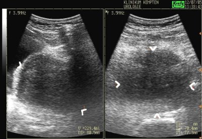
After micturition, the bladder's length, width, and height [in cm] are recorded in sagittal and horizontal planes [fig. postvoid residual volume]. The postvoid residual volume is calculated using the formula:
Postvoid residual volume (PVR) = length × width × height × 0.5
Various factors ranging from 0.5–0.7 are found in the literature. Depending on the bladder shape, considerable inaccuracy is possible. Usually, there is no residual urine. PVR volumes over 100–ml are considered significant in adults. In children, a PVR volume exceeding 10% of the urinary bladder capacity is pathological (Riedmiller et al., 2001). Please see the following table for the differential diagnosis of PVR.
| DD urinary retention | |
| Diseases of the prostate | Benign prostatic hyperplasia, prostate cancer, bladder cancer, bladder stones, bladder neck sclerosis, detrusor sphincter dyssynergia (DSD). |
| Diseases of the urethra | Urethral strictures, urethral valves, female urethral cancer and male urethral cancer, foreign bodies, phimosis. |
| Diseases of the urinary bladder | Neurogenic lower urinary tract dysfunction, drugs (anticholinergic drugs, neuroleptics), pelvic surgery, diabetes mellitus. |
Transabdominal Ultrasonography of the Prostate
Transabdominal imaging of prostate is possible through the filled bladder. After determining the length, width, and height in two planes, the prostate volume is calculated using the formula:
Prostate volume = Length × Width × Height × 0.5.
The transabdominal measurement method offers advantages in huge prostate glands due to the larger field of vision [fig. transabdominal prostate ultrasound]. In addition, transabdominal ultrasonography of the prostate quickly identifies enlarged median lobes or prostatic cysts.
 |
| Ureter ultrasound | Index | Penis ultrasound |
Index: 1–9 A B C D E F G H I J K L M N O P Q R S T U V W X Y Z
References
Singer u.a. 2006 SINGER, Eric A. ; GOLIJANIN,
Dragan J. ; DAVIS, Robert S. ; DOGRA, Vikram:
What’s new in urologic ultrasound?
In: Urol Clin North Am
33 (2006), Aug, Nr. 3, S. 279–286
 Deutsche Version: Sonographie der Harnblase und Restharnmessung
Urology-Textbook.com – Choose the Ad-Free, Professional Resource
This website is designed for physicians and medical professionals. It presents diseases of the genital organs through detailed text and images. Some content may not be suitable for children or sensitive readers. Many illustrations are available exclusively to Steady members. Are you a physician and interested in supporting this project? Join Steady to unlock full access to all images and enjoy an ad-free experience. Try it free for 7 days—no obligation.
New release: The first edition of the Urology Textbook as an e-book—ideal for offline reading and quick reference. With over 1300 pages and hundreds of illustrations, it’s the perfect companion for residents and medical students. After your 7-day trial has ended, you will receive a download link for your exclusive e-book.