You are here: Urology Textbook > Bladder > Neurogenic Lower Urinary Tract Dysfunction
Neurogenic Lower Urinary Tract Dysfunction: Etiology and Symptoms
Neurogenic lower urinary tract dysfunction (NLUTD) refers to abnormal function of the bladder, bladder neck, and urinary sphincters related to a neurologic disorder. Old terminology: neurogenic bladder. (Chapple u.a., 2005) (van Kerrebroeck, 1998) (Wein und Rackley, 2006). EAU Leitlinie: NLUTD.
Classification of NLUTD
Simplified classification:
The function of the lower urinary tract is divided into the storage and emptying phases. Neurogenic dysfunction of the storage or emptying phase is caused by a disorder of the bladder function (detrusor), the urethral function (sphincter), or a combination.
International Continence Society:
The function of the lower urinary tract is divided into the storage and emptying phases; further classification depends on the urodynamic findings, see table ICS classification of NLUTD.
Clinically Defined Disorders
Overactive bladder (OAB):
OAB is urinary urgency, with or without urgency incontinence, usually with increased daytime frequency and nocturia, if there is no proven infection or other obvious pathology (Abrams et al., 2002). Other synonyms are urge syndrome or urgency-frequency syndrome; see section overactive bladder for details.
Urge incontinence:
Urge incontinence is the involuntary loss of urine, accompanied by a sudden, compelling desire to void.
Stress urinary incontinence:
Stress urinary incontinence is the involuntary loss of urine on effort or physical exertion, like sporting activities, sneezing, or coughing.
Neurogenic Lower Urinary Tract Dysfunctions Defined by Urodynamics
The following disorders can only be differentiated using urodynamic studies; see also table ICS classification of NLUTD.
Detrusor overactivity (DO):
Detrusor overactivity may be phasic (detrusor pressure increase during bladder filling without complete emptying of the bladder) or terminal (detrusor contraction during bladder filling with complete emptying of the bladder). Neurogenic DO may be attributed to a neurological disease; the older term is detrusor hyperreflexia. The cause for idiopathic DO remains unclear (older term detrusor instability).
Detrusor sphincter dyssynergia (DSD):
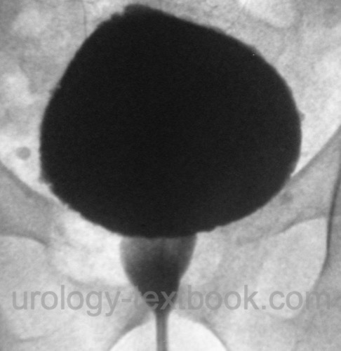
Detrusor sphincter dyssynergia (DSD) is defined by a detrusor contraction with simultaneous involuntary pelvic floor activation caused by a neurological disease [fig. signs of DSD in VCUG]. Classic DSD only occurs in lesions between the pontine and sacral micturition center (e.g., spinal cord injury). The DSD leads to high detrusor leak point pressures (DLPP) of the urinary bladder. Without therapy, 50% of patients with DSD develop upper urinary tract complications.
Dysfunctional voiding:
Dysfunctional voiding is characterized by an intermittent urine stream due to intermittent pelvic floor contractions in the EMG in patients with no recognizable neurological disease. Synonyms: idiopathic detrusor-sphincter dyssynergia or detrusor-sphincter dyscoordination.
 |
Epidemiology of NLUTD
In children, the prevalence of neurogenic lower urinary tract dysfunction is rare; the most common causes are spina bifida, spinal cord injury, cerebral palsy, and anorectal malformations. The prevalence of NLUTD increases with age due to the epidemiology of the underlying neurological diseases.
Pathogenesis, Signs, and Symptoms of Neurogenic Lower Urinary Tract Dysfunction
General principles:
Neurological lesions above the brainstem lead to detrusor overactivity of variable extent with possibly sphincter dyssynergia. The sphincter control is mostly preserved.
Complete spinal cord injuries below Th6 initially lead to spinal shock with atonic urinary bladder, then to detrusor overactivity with detrusor-sphincter dyssynergia of the striated sphincter muscle. Spinal cord injuries over Th6 also lead to smooth muscle sphincter dyssynergia and autonomic hyperreflexia.
Sacral lesions and lesions of the peripheral reflex arc lead to a hypocontractile detrusor, increased urinary bladder capacity and variable residual sphincter activity without dyssynergia.
Cerebrovascular accident (Stroke, CVA):
Acute CVA leads to a hypocontractile detrusor and urinary retention. Detrusor overactivity with a preserved sphincter function develops after several weeks. The prevalence of incontinence after CVA is 20–30% in the long term; the causes are detrusor overactivity, reduced control of the striated sphincter, and the reduced perception of bladder filling.
Traumatic brain injury:
Signs and symptoms similar to that after CVA, injury of the brain stem may lead to DSD.
Dementia:
Incontinence may be caused by intellectual deficits or detrusor overactivity. Anticholinergics are contraindicated in Alzheimer disease.
Parkinson disease:
Parkinson disease causes voiding disorders in 35–70%, mostly detrusor overactivity with urge incontinence. In addition, a lack of control over the sphincter and a detrusor weakness is possible. Surgery for bladder outlet obstruction should be done with caution and may lead to inferior results as usually expected.
Central system atrophies:
Shy-Drager syndrome and olivopontocerebellar atrophy are neurological diseases with extrapyramidal, cerebellar, pyramidal, and autonomic failure symptoms. Voiding symptoms (incontinence, urge) often precede the diagnosis by years.
Multiple sclerosis (MS):
MS is a chronic demyelinating disease of the CNS of unknown etiology with ataxia, nystagmus, paralysis, and neurogenic lower urinary tract dysfunction (50–90%). Voiding symptoms are the first in 10% of patients with MS, usually urinary retention or urgency. Depending on the location of the lesions, urodynamics may reveal detrusor overactivity (30–90%), DSD (30–65%), or hypocontractile detrusor (5–20%).
Spinal Cord Injury:
After acute spinal cord injury, flaccid paresis of the affected muscle groups and an absent sensation of the dermatomes below the lesion develop. In addition, peripheral vasodilation and spinal shock may develop. Acute injury leads to an acontractile detrusor with urinary retention since the smooth sphincter compensates for the weakness of the striated bladder sphincter. The phase of hypocontractile detrusor is followed by the development of neurogenic detrusor overactivity after 6–12 weeks; see the following section. Detrusor overactivity may develop within a few days if the lesion is incomplete.
Lesions of the spinal cord above the sacral micturition center:
Possible causes of lesions above the sacral micturition center are trauma above the first lumbar vertebra, multiple sclerosis, infarcts, infections, and tumors. The sacral micturition reflex remains intact; the pontine and cortical inhibitions are eliminated. The lack of central inhibition results in neurogenic detrusor overactivity and detrusor-sphincter dyssynergia. Patients develop a bladder with reduced functional capacity, high intravesical micturition pressures, bladder wall hypertrophy, spasticity of the pelvic floor muscles, and the risk of damage to the upper urinary tract due to reflux and hydronephrosis. The extent of detrusor overactivity and DSD varies significantly in individual cases, depending on the location and extent of the neurological lesion.
Autonomic dysreflexia:
Autonomic dysreflexia is a potentially fatal emergency in patients with paraplegia. Manipulation of the urinary tract can trigger a reaction of the sympathetic nervous system (autonomic dysreflexia) in patients with lesions above Th5. Signs and symptoms are bradycardia, hypertensive crises, headaches, piloerection, and sweating. Autonomous dysreflexia requires an emergency treatment to lower the blood pressure. Prophylaxis with 10 to 20 mg nifedipine s.l. is possible. In severe cases, sacral deafferentation is necessary.
Lesions of the sacral spinal cord:
Causes are trauma below L1, infections with polio or herpes zoster, herniated discs, radiation therapy, operations, tumors, infarcts, or spina bifida. Sacral spinal cord damage leads to a hypocontractile or acontractile detrusor. Since the lesion is rarely complete, mixed symptoms with detrusor overactivity may occur. The external sphincter tonus is reduced, but urinary incontinence is not mandatory due to smooth muscle sphincter tonus and compensatory bladder enlargement.
Cauda equina syndrome:
Cauda equina syndrome consists of saddle anesthesia, erectile dysfunction, and loss of voluntary control over the anal and urinary bladder sphincter due to cauda equina lesions.
Lesions of the afferent innervation:
Causes are diabetes mellitus, syphilis (tabes dorsalis), pernicious anemia, genital herpes, or dorsal horn lesions of the spinal cord. The sensation of bladder filling is impaired, resulting in an overdistension of the urinary bladder with subsequent myogenic detrusor insufficiency and risk of urinary retention.
Diabetes mellitus:
Peripheral neuropathy leads to an impaired afferent and efferent innervation of the lower urinary tract, leading to a gradual increase in urinary bladder capacity and impaired detrusor function with the risk of urinary retention. Early diagnosis and institution of timed voiding can prevent chronic bladder distention in most cases.
Lesions of the peripheral nerves:
Causes are radical rectum surgery, radical hysterectomy, pelvic fractures, and radiation. The loss of the sacral micturition reflex leads to a hypocontractile (areflexive) urinary bladder with urinary retention since the striated sphincter function is not impaired. Bladder filling is impaired (decreased compliance) since the detrusor is no longer inhibited in response to bladder filling.
| Overactive Bladder | Index | NLUTD Treatment |
Index: 1–9 A B C D E F G H I J K L M N O P Q R S T U V W X Y Z
References
Abrams, P.; Cardozo, L.; Fall, M.; Griffiths, D.;
Rosier, P.; Ulmsten, U.; van Kerrebroeck, P.; Victor, A.; Wein, A. & of
the International Continence Society, S. S.
The standardisation of
terminology of lower urinary tract function: report from the
Standardisation Sub-committee of the International Continence Society.
Neurourol
Urodyn, 2002, 21, 167–178.
A. W. Partin, C. A. Peters, L. R. Kavoussi, R. R. Dmochowski, and A. J. Wein, Campbell-Walsh-Wein Urology, 12th ed. ISBN-13: 978-1455775675: Elsevier, 2020.
Chapple u.a. 2005 CHAPPLE, C. ; KHULLAR, V. ;
GABRIEL, Z. ; DOOLEY, J. A.:
The effects of antimuscarinic treatments in overactive bladder: a
systematic review and meta-analysis.
In: Eur Urol
48 (2005), Nr. 1, S. 5–26
van Kerrebroeck 1998 KERREBROECK, P. E. V. van:
Neurogenic Bladder Dysfunction.
In: Eur Urol
Curric Urol 4.2 (1998), S. 1–9
Wein und Rackley 2006 WEIN, A. J. ; RACKLEY,
R. R.:
Overactive bladder: a better understanding of pathophysiology,
diagnosis and management.
In: J Urol
175 (2006), Nr. 3 Pt 2, S. S5–10
 Deutsche Version: Neurogene Funktionsstörungen des unteren Harntrakts
Urology-Textbook.com – Choose the Ad-Free, Professional Resource
This website is designed for physicians and medical professionals. It presents diseases of the genital organs through detailed text and images. Some content may not be suitable for children or sensitive readers. Many illustrations are available exclusively to Steady members. Are you a physician and interested in supporting this project? Join Steady to unlock full access to all images and enjoy an ad-free experience. Try it free for 7 days—no obligation.
New release: The first edition of the Urology Textbook as an e-book—ideal for offline reading and quick reference. With over 1300 pages and hundreds of illustrations, it’s the perfect companion for residents and medical students. After your 7-day trial has ended, you will receive a download link for your exclusive e-book.