You are here: Urology Textbook > Urologic examinations > Imaging > Penis ultrasound
Penis Ultrasound Imaging and Doppler Sonography
Penile ultrasound is used in the diagnostic workup of a patient with erectile dysfunction, Peyronie’s disease, priapism, penile fracture, penile urethral strictures, urethral stones, urethral diverticulae and penile cancer involving deep tissues of the penis. Penile Doppler ultrasound is used in addition in patients with erectile dysfunction and priapism.
 |
 |
Examination Technique
A high-resolution linear probe of 7.5–10 MHz is used for imaging of the penis. The penis is examined longitudinally and in transversal sections with the probe on the dorsum and/or ventrum of the penis [fig. transverse ultrasound imaging from ventral and transverse ultrasound imaging from dorsal]. The tunica albuginea is a thin echogenic structure surrounding the hypoechogenic erectile tissue. The penile vessels can be identified dorsal to the tunica albuginea. The lumen of the urethra cannot be seen without injection of lubrication gel, after the injection the urethra is examined from the ventrum of the penis. Imaging of the urethra is best done with an assistant who stretches the penis to the side while injecting the lubricant, this enables the examiner to do imaging with both hands free [see section urethral stricture].
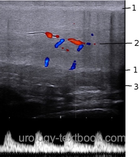
Penile Doppler Ultrasonography
The measurement of penile blood flow after pharmacologically induced erection provides valuable information about the erectile function, see section diagnostic workup of erectile dysfunction for details. The quality of erection is assessed every 5–10 min after intracavernous injection of alprostadil until full erection is achieved and a Doppler sonography of the penile arteries and veins is done. If the erection does not occur within 30 minutes, the examination will be terminated.
Measurements of Doppler sonography are done at the cavernous artery in the corpus cavernosum [fig. Penile Doppler ultrasonography (longitudinal section)]. The standard value for the peak systolic velocity (PSV) is at least 25 cm/s. At full erection, the end-diastolic velocity (EDV) in the arteries should be below 5 cm/s, higher values suggest venous leakage from the corpus cavernosum. The flow in the penile veins should also be below 5 cm/s. The RI (resistive index) is close to 1 at maximum erection because end-diastolic blood flow should near to zero. An RI below 0.75–0.8 indicates venous leakage.
 |
| Bladder ultrasound | Index | Testicular ultrasound |
Index: 1–9 A B C D E F G H I J K L M N O P Q R S T U V W X Y Z
References
Singer u.a. 2006 SINGER, Eric A. ; GOLIJANIN, Dragan J. ; DAVIS, Robert S. ; DOGRA, Vikram: What’s new in urologic ultrasound?In: Urol Clin North Am
33 (2006), Aug, Nr. 3, S. 279–286
 Deutsche Version: Sonographie des Penis
Urology-Textbook.com – Choose the Ad-Free, Professional Resource
This website is designed for physicians and medical professionals. It presents diseases of the genital organs through detailed text and images. Some content may not be suitable for children or sensitive readers. Many illustrations are available exclusively to Steady members. Are you a physician and interested in supporting this project? Join Steady to unlock full access to all images and enjoy an ad-free experience. Try it free for 7 days—no obligation.
New release: The first edition of the Urology Textbook as an e-book—ideal for offline reading and quick reference. With over 1300 pages and hundreds of illustrations, it’s the perfect companion for residents and medical students. After your 7-day trial has ended, you will receive a download link for your exclusive e-book.