You are here: Urology Textbook > Kidneys > Autosomal dominant polycystic kidney disease
Autosomal Dominant Polycystic Kidney Disease (ADPKD)
Definition of Autosomal Dominant Polycystic Kidney Disease
Autosomal dominant polycystic kidney disease is an inherited cystic renal disease with the development of end-stage renal disease in adulthood (Kuehn and Walz, 2007).
Epidemiology
- Incidence 1:2000
- 15% of dialysis patients are suffering from ADPKD
Etiology of Autosomal Dominant Polycystic Kidney Disease
Genetics:
Variations in two genes are known to cause aut. dom. polycystic kidney disease. In 85–90% of patients with ADPKD, mutations in PKD1 (chromosome 16, coding for polycystin-1) are responsible for the disease. In 10–15%, mutations of PKD2 (chromosome 4, coding for polycystin-2) are responsible. ADPKD caused by mutations of PKD2 usually has a later onset and slower progression of the disease. Autosomal dominant inheritance with almost 100% penetrance is typical, so 50% of children will inherit the disease from affected patients. ADPKD occurs to Knudson's theory of two hits: one diseased gene is inherited, and the second copy of the gene is damaged by a spontaneous mutation. This explains the long asymptomatic latency in the onset of the disease.
Pathophysiology:
Polycystin-1 and polycystin-2 have essential functions in signal transduction and in forming the primary cilium of the tubular epithelial cells. Defective polycystin leads to the tubular epithelium's proliferation and cyst formation; every part of the nephron may be affected. Similar mechanisms may damage blood vessels and other organ systems. The disease leads to the activation of mTOR signaling, enhances cAMP production, and increases ion and fluid secretion into the renal cystic lumen. The vasopressin V2-receptor blocker tolvaptan inhibits ADH dependent cAMP production, slows the increase in kidney volume, and delays the development of chronic kidney disease.
Progression factors:
The normal (bilateral) kidney volume is usually below 400 ml. In ADPKD, increasing renal volume is associated with decreasing renal function. The kidney volume can be measured either by MRI or renal ultrasound imaging: the greater the kidney volume, the worse the prognosis for renal function. The time to end-stage renal disease for a 30-year-old man depends on the renal volume: 10 years (renal volume 2000 ml), 18 years (1500 ml) or 21 years (1000 ml) (Kuehn et al., 2015).
Pathology:
Autosomal dominant polycystic kidney disease leads to enlarged kidneys with multiple cysts [fig. polycystic kidneys]. The cysts are a few millimeters to a centimeter in size and derive from tubules of the nephron. The epithelium of the cyst corresponds to its origin. Cysts also develop in other organ systems; see section signs and symptoms [fig. liver cysts].
| Do you want to see the illustration? Please support this website with a Steady membership. In return, you will get access to all images and eliminate the advertisements. Please note: some medical illustrations in urology can be disturbing, shocking, or disgusting for non-specialists. Click here for more information. |
| Do you want to see the illustration? Please support this website with a Steady membership. In return, you will get access to all images and eliminate the advertisements. Please note: some medical illustrations in urology can be disturbing, shocking, or disgusting for non-specialists. Click here for more information. |
| Do you want to see the illustration? Please support this website with a Steady membership. In return, you will get access to all images and eliminate the advertisements. Please note: some medical illustrations in urology can be disturbing, shocking, or disgusting for non-specialists. Click here for more information. |
Signs and Symptoms
Onset of disease:
Symptoms start at the age of 30 to 50 years. Renal ultrasound and genetic screening lower the average age of initial diagnosis due to the discovery of the asymptomatic disease. Rarely the disease begins in infancy.
Symptoms:
- Hematuria (50%)
- Proteinuria
- Flank pain and fever (cyst rupture or cyst infection)
- Nephrolithiasis
- Gastrointestinal symptoms due to large kidneys
- Arterial hypertension (80%)
- End-stage renal disease and symptoms of uremia in the fifth decade (PDK1 mutation) or seventh decade (PDK2 mutation)
Manifestations in other organs:
- Cysts in the liver, pancreas, spleen, and lungs
- Aneurysm of the cerebral arteries
- Colonic diverticula
- Mitral valve prolapse
Diagnostic Workup of Autosomal Dominant Polycystic Kidney Disease
Family history:
Covering at least three generations.
Laboratory examinations:
- Urine analysis for proteinuria
- Creatinine or/and cystatin C to evaluate renal function.
- Genetic evaluation: for the differential diagnosis of unclear cystic kidney disease and as a predictive diagnosis in case of a positive family history. A parent with ADPKD has a 50% chance of having a child with the same condition (autosomal dominant inheritance pattern). Genetic predictive testing is costly and offers few (if any) clinical advantages compared to screening with imaging (see the following paragraph).
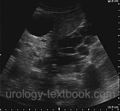
Renal ultrasound:
Renal imaging enables an early diagnosis of patients at risk (with a positive family history). The diagnosis of ADPKD is certain in patients at risk if there are at least two unilateral renal cysts or bilateral renal cysts at an age below 30 years, or at least two renal cysts in each kidney (30–60 years old). Further ultrasound imaging should focus on the liver, pancreas, and spleen. If cysts are not detectable in the kidneys or visceral organs at 40 years, a patient at risk is considered healthy (Ravine et al., 1994). The determination of the bilateral renal volume is important for affected patients to evaluate the prognosis of the renal function (see above). Later in the disease, the kidneys are massively enlarged and filled with cysts.
 |
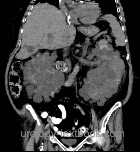
CT or MRI:
- Abdominal imaging: in patients with flank pain or suspect ultrasound imaging.
- Cranial imaging: to estimate the risk of intracranial bleeding due to aneurysms.
 |
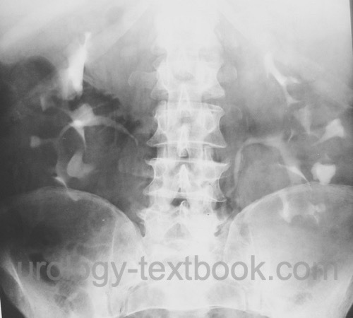
Intravenous urography:
Intravenous urography is only indicated, if abdominal CT is not available. Urography reveals bilateral renal enlargement, calyceal stretching from the cysts, and Swiss cheese aspect in the nephrographic phase.
 |
Treatment of Autosomal Dominant Polycystic Kidney Disease
Medical Treatment:
Early therapy of arterial hypertension with ACE inhibitors or angiotensin-II receptor antagonists. Low-salt diet. Protein reduction does not improve the prognosis. The vasopressin (V2) receptor antagonist tolvaptan has been approved in patients at high risk of progression (age <30 years, renal volume >1500 ml and GFR <90 ml) to slow down disease progression (Torres et al., 2012). It is realistic to postpone dialysis for 4–6.5 years. Polyuria (sometimes over 7 l/d) and idiosyncratic hepatotoxicity are problematic side effects of tolvaptan that require intensive patient monitoring.
Management of Flank Pain:
Nephrolithiasis, bleeding, or infection of cysts may be responsible for flank pain. If imaging can reveal altered cysts, percutaneous management (cyst aspiration and sclerotherapy) or laparoscopic deroofing of cysts may be helpful.
Treatment of Renal Failure:
Hemodialysis and kidney transplantation are the treatment alternatives. Nephrectomy is often necessary before renal transplantation: either to create room for the transplant or if recurrent symptoms from the enlarged polycystic kidneys are present.
Experimental Treatment Approaches:
Limitation of tubulus epithelium proliferation by inhibitors of signal transduction. Previous studies with mTOR inhibitors, such as everolimus, have failed to demonstrate any protective effect on renal function. Prospects include EGFR tyrosine kinase inhibitors or somatostatin analogs (Walz et al., 2010).
Prognosis of Autosomal Dominant Polycystic Kidney Disease (ADPKD):
The prognosis has improved due to better treatment options for complications like urinary tract infection, kidney stones, hypertension or chronic kidney disease.
Renal failure:
ADPKD leads to dialysis in 2% by the age of 40, 23% by the age of 50, and 48% by the age of 73. The risk for end-stage renal disease correlates closely with kidney volume (see above).
Cerebral hemorrhage:
9% of patients with ADPKD die of subarachnoid hemorrhage (ruptured brain aneurysm). In addition, cerebral hemorrhage due to malignant hypertension is possible.
| ARPKD | Index | Kidney diseases |
Index: 1–9 A B C D E F G H I J K L M N O P Q R S T U V W X Y Z
References
W. Kuhn and G. Walz. Autosomal dominante polyzystische nierenerkrankung.Dtsch Arztebl, 104 (44): 3022–8, 2007.
Walz, G.; Budde, K.; Mannaa, M.; Nürnberger, J.; Wanner, C.; Sommerer, C.; Kunzendorf, U.; Banas, B.; Hörl, W. H.; Obermüller, N.; Arns, W.; Pavenstädt, H.; Gaedeke, J.; Büchert, M.; May, C.; Gschaidmeier, H.; Kramer, S. & Eckardt, K. Everolimus in patients with autosomal dominant polycystic kidney disease.
N Engl J Med, 2010, 363, 830-840
 Deutsche Version: ADPKD
Urology-Textbook.com – Choose the Ad-Free, Professional Resource
This website is designed for physicians and medical professionals. It presents diseases of the genital organs through detailed text and images. Some content may not be suitable for children or sensitive readers. Many illustrations are available exclusively to Steady members. Are you a physician and interested in supporting this project? Join Steady to unlock full access to all images and enjoy an ad-free experience. Try it free for 7 days—no obligation.
New release: The first edition of the Urology Textbook as an e-book—ideal for offline reading and quick reference. With over 1300 pages and hundreds of illustrations, it’s the perfect companion for residents and medical students. After your 7-day trial has ended, you will receive a download link for your exclusive e-book.