You are here: Urology Textbook > Anatomy > Kidney > Histology of the renal tubules
Histology of the kidney (3/7): Renal Tubules
- Anatomy of the kidney (1/7): Gross anatomy
- Anatomy of the kidney (2/7): Histology of the glomerulus and nephron
- Anatomy of the kidney (3/7): Histology of renal tubules
- Anatomy of the kidney (4/7): Physiology of the glomerular filtration rate
- Anatomy of the kidney (5/7): Physiology of the tubular reabsorption
- Anatomy of the kidney (6/7): Physiology of the renin-angiotensin-aldosterone system
- Anatomy of the kidney (7/7): Physiology of erythropoetin, endothelins and vitamin D
Review literature: (Benninghoff, 1993).
 |
Proximal Tubule of the Kidney
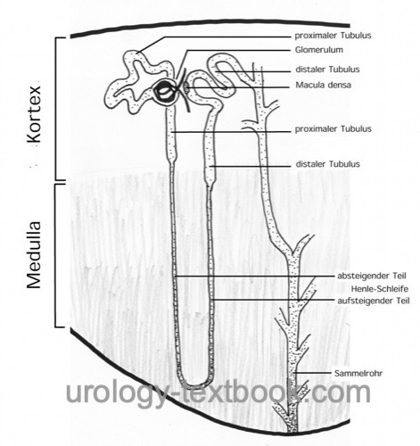
The proximal tubule of the kidney has two parts [schematic figure of the nephron]:
- Proximal Convoluted Tubule (PCT): the pars convoluta is the first segment of the renal tubules and gets the primary urine of the glomerulus
- Proximal Straight Tubule (PST): the pars recta of the proximal tubule leads the urine to the first part of the Henle loop.
Both parts of the proximal tubule have a low columnar epithelium with a wide brush border and strong basolateral folds (as seen in light microscopy with basal striations). The basal cell compartment is rich of mitochondria; this enables active transport processes with the help of the Na-K-ATPase.
Loop of Henle
The loop of Henle has two different loop lengths: a short loop, which descends to the cortical-medullary border, and a long loop, which descends to the renal papilla. Furthermore, there are three different parts of the loop of Henle:
- Descending Thin Limb (DTL)
- Ascending Thin Limb (ATL)
- Thick Ascending Limb (TAL)
The short Henle loops have a descending thin limb and a thick ascending limb. The long Henle loops have a thin descending and ascending thin limb, then the pars recta of the distal tubule follows.
The thin parts of the loop of Henle are lined with a single-layer flat epithelium with a high permeability for water (and ions via solvent drag) through a weak tight junction (zonula occludens).
The thick ascending limb of the Henle loop is lined by low cuboidal cells without a brush border. The epithelium cannot be penetrated by water.
Distal Tubule of the Kidney
The distal tubule of the kidney has three parts [schematic figure of the nephron]:
- Thick Ascending Limb (TAL): also called pars recta of the distal tubule.
- Macula densa: specialized cells of the distal tubule, which are in contact with the mesangial cells of the glomerulus.
- Distal Convoluted Tubule (DCT): the pars convoluta of the distal tubule
The distal tubule is lined with by low cuboidal cells without a brush border. The epithelium shows the typical structure suitable for high absorptive functions (microvilli, basolateral membrane folds with abundant mitochondria).
Macula densa and Juxtaglomerular Apparatus
Specialized cells form the macula densa of the distal tubule, which contact with the basal side the mesangial cells of the glomerulus. The macula densa is an epithelium with resorptive capacities to basal, where the mesangial cells with receptor functions can analyze the urine. Macula densa, mesangial cells and renin-producing cells of the afferent arterioles form together the juxtaglomerular apparatus. With the aid of the juxtaglomerular apparatus, each nephron has the possibility to change the filtration performance in the glomerulus, depending on the urine analysis in the distal tubule.
Collecting Duct
Collecting Ducts (CD) transport the urine via the renal papillae into the renal calyx [schematic figure of the nephron]. Collecting ducts are lined with cuboidal epithelium containing two cell types: the principle cells (bright staining cytoplasm) and intercalated cells (dark staining cytoplasm).
The intercalated cells are scattered in the epithelium and participate in acid-base homeostasis. Type A intercalated cells are specialized for proton secretion. A defect of the type A cells leads to distal renal tubular acidosis. Type B intercalated cells secrete bicarbonate. Most cells of the epithelium are principal cells, which regulate the sodium and potassium concentrations under control of aldosterone and ADH.
| Nephron (Glomerulus) | Index | Glomerular filtration |
Index: 1–9 A B C D E F G H I J K L M N O P Q R S T U V W X Y Z
References
Benninghoff, A. (1993): Makroskopische Anatomie, Embryologie und Histologie des Menschen. 15. Auflage. München; Wien; Baltimore: Urban & Schwarzenberg.
 Deutsche Version: Histology der Nieren: Tubuli (distales Nephron).
Urology-Textbook.com – Choose the Ad-Free, Professional Resource
This website is designed for physicians and medical professionals. It presents diseases of the genital organs through detailed text and images. Some content may not be suitable for children or sensitive readers. Many illustrations are available exclusively to Steady members. Are you a physician and interested in supporting this project? Join Steady to unlock full access to all images and enjoy an ad-free experience. Try it free for 7 days—no obligation.
New release: The first edition of the Urology Textbook as an e-book—ideal for offline reading and quick reference. With over 1300 pages and hundreds of illustrations, it’s the perfect companion for residents and medical students. After your 7-day trial has ended, you will receive a download link for your exclusive e-book.