You are here: Urology Textbook > Anatomy > Kidney > Gross anatomy
Kidney: Gross Anatomy, Renal Fascia, Vessels, and Nerves
- Anatomy of the kidney (1/7): Gross anatomy
- Anatomy of the kidney (2/8): Histology of glomerulus and nephron
- Anatomy of the kidney (3/7): Histology of renal tubules
- Anatomy of the kidney (4/7): Physiology of the glomerular filtration rate
- Anatomy of the kidney (5/7): Physiology of the tubular reabsorption
- Anatomy of the kidney (6/7): Physiology of the renin-angiotensin-aldosterone system
- Anatomy of the kidney (7/7): Physiology of erythropoetin, endothelins and vitamin D
Review literature: (Benninghoff, 1993).
Gross Anatomy of the Kidney
The kidneys are paired organs; one kidney weighs 120–200 g. The normal kidney measures 10–12 cm vertically, 5–7 cm transversely, and 3 cm a.p. The outer surface can be divided into anterior side, dorsal side, upper pole, and lower pole. The lateral surface is convex. The medial surface is concave, deeply grooved (renal sinus), and forms the renal hilum. The ureter/renal pelvis, renal artery, renal vein, lymphatic vessels, and nerves enter or exit through the renal hilum of the kidney.
Renal Fascias
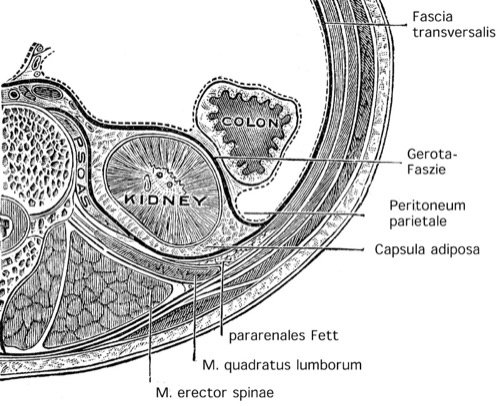
The kidneys have a tough organ capsule (Capsula fibrosa), which follows medially the renal parenchyma and does not surround the renal hilum. The kidneys are surrounded by a layer of perinephric fat (Capsula adiposa). A connective tissue sheath, fascia renalis or Gerota fascia, surrounds the kidney with perinephric fat and the adrenal gland. Cranially and laterally, the renal fascia is closed by fusion of the anterior and posterior sheath. Medially and inferiorly, there is no connection between the anterior and posterior sheath of the renal fascia: renal fluid collections can drain into the pelvis. The anterior sheath of the renal fascia lies immediately below the parietal peritoneum [fig . renal and retroperitoneal fascias].
 |
Topographic Anatomy of the Kidneys
The kidneys are located in the retroperitoneum, right and left of the spine, and below the diaphragm. Normal position:
- Left kidney: from the 11th rib to the third lumbar vertebra.
- Right kidney: slightly lower position, 12th rib to the lower part of the third lumbar vertebra.
Topographic Anatomy of the Right Kidney
- Dorsally: 12th rib, subcostal nerve, iliohypogastric nerve, ilioinguinal nerve, quadratus lumborum muscle, diaphragm with pleura at the upper pole
- Cranially: adrenal gland.
- Medially: psoas major muscle, inferior vena cava, ovarian/testicular vein, ureter.
- Ventrally: liver, duodenum, ascending colon.
Topographic Anatomy of the Left Kidney
- Dorsally: 11th and 12th rib, subcostal nerve, iliohypogastric nerve, ilioinguinal nerve, quadratus lumborum muscle, diaphragm with pleura at the upper pole
- Cranially: adrenal gland, spleen.
- Medially: psoas major muscle, aorta, ovarian/testicular vein, ureter.
- Ventrally: spleen, pancreas, stomach, descending colon.
Internal Anatomy of the Kidneys
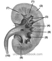
The renal parenchyma has two distinct components: the renal cortex and the renal medulla. Since the form of the renal medulla is a pyramid shape, it is also called a renal pyramid. In the longitudinal section, the individual functional units of the kidney can be seen [fig. internal anatomy of the kidney]. The kidney consists on average of 14 renal lobes. Each lobe has a renal pyramid and is surrounding by cortex tissue. Each lobe drains with its collecting tubules to its renal calyx; the collecting tubules indent the calyx and form the renal papilla. Each kidney has 7–13 calyces, some lobes drain together in a single calyx. The urine drains from the calyx via the renal pelvis into the ureter. The cortex-like parenchyma between the renal pyramids is called the renal columns of Bertin (Columnae renales).
During embryonic development, the morphological separation of the individual renal lobes is clearly visible, with deep incisions separating the individual renal lobes (renkulation). The surface of the kidneys smooths out during the first few years of life. Occasionally, renkulation persists into adulthood.
 |
Vessels of the Kidney: Renal Artery, Vein and Nerves
Renal Artery
The kidneys are supplied by paired branches of the aorta. The vascular supply (A. renalis dexter et sinistra) is often subject to variations. The right renal artery passes dorsal to the vena cava to the right kidney. Both renal arteries run together with the renal veins.
Renal Vein
The renal veins drain into the inferior vena cava. The left renal vein is longer and passes over the aorta below the superior mesenteric artery into the vena cava.
Lymphatic Vessels of the Kidney
The left kidney drains into the paraaortic lymph nodes. The right kidney drains into the paracaval and interaortocaval lymph nodes. The cranial and caudal extent of the renal lymph nodes along the vena cava and aorta ranges from the inferior mesenteric artery to the diaphragm.
Innervation of the kidney
The renal plexus is located around the renal artery and contains postganglionic fibers from the sympathetic nervous system (Th10 to L2). The nerve fibers from the plexus enter the kidney with the branches of the renal artery and regulate the vascular tone and the secretion of renin.
The renal function is not dependent on the innervation mentioned above, as shown with the renal function after kidney transplantation (complete transection of the innervation). Hormones regulate most renal functions.
| Anatomy | Index | Kidney: nephron |
Index: 1–9 A B C D E F G H I J K L M N O P Q R S T U V W X Y Z
References
Benninghoff, A. (1993): Makroskopische Anatomie, Embryologie und Histologie des Menschen. 15. Auflage. München; Wien; Baltimore: Urban & Schwarzenberg.
 Deutsche Version: Anatomie der Nieren.
Urology-Textbook.com – Choose the Ad-Free, Professional Resource
This website is designed for physicians and medical professionals. It presents diseases of the genital organs through detailed text and images. Some content may not be suitable for children or sensitive readers. Many illustrations are available exclusively to Steady members. Are you a physician and interested in supporting this project? Join Steady to unlock full access to all images and enjoy an ad-free experience. Try it free for 7 days—no obligation.
New release: The first edition of the Urology Textbook as an e-book—ideal for offline reading and quick reference. With over 1300 pages and hundreds of illustrations, it’s the perfect companion for residents and medical students. After your 7-day trial has ended, you will receive a download link for your exclusive e-book.