You are here: Urology Textbook > Ureters > Ureterocele
Ureterocele: Symptoms, Diagnosis and Treatment
Definition
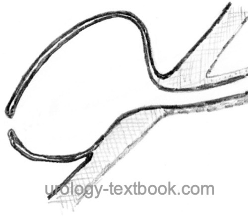
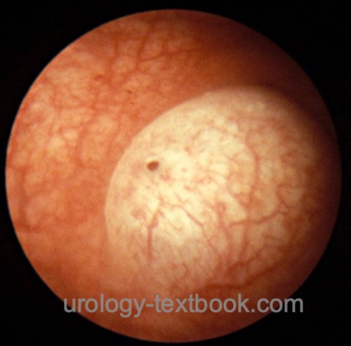
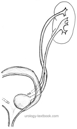
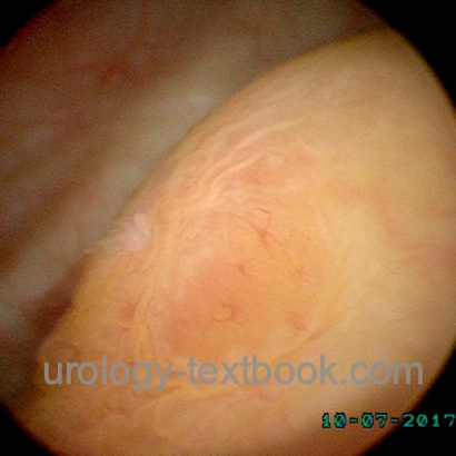
The ureterocele is a cystic dilatation of the distal, intravesical ureter [fig. ureterocele cross-section and ureterocele in cystoscopy]. In 80%, the ureterocele drains the upper pole of a duplex kidney [fig. ureterocele with duplex kidney]. A ureterocele causes a defect in the trigonum and predisposes the patient to vesicoureteral reflux into the ureter of the lower pole of a duplex kidney. EAU guideline: Paediatric Urology.
 |
 |
The following classifications exist:
- By Ericsson: separates between intravesical (simple) ureterocele from ectopic ureteroceles, which extend to the bladder neck, and urethra.
- By Stephens:
- Intravesical ureteroceles: which may be stenotic or non-obstructed.
- Extravesical ureteroceles: which may be sphincteric (non-obstructive ostium distal of the bladder neck), sphincterostenotic (stenotic ostium distal of the bladder neck), caecoureterocele (intravesical ostium, but the ureterocele extends down into the urethra and causes obstruction) or blind (no contact to the kidney).
 |
Epidemiology of Ureterocele
Prevalence 1:5000, more often in females (4:1).
Etiology of Ureterocele
Different theories exist, but no theory can explain all forms of ureteroceles:
Chwalla membrane:
The Chwalla membrane separates the urogenital sinus from the ureteral bud. A delayed and incomplete reabsorption of Chwalla membrane is postulated, causing a stenotic ostium which also leads to the cystic dilatation of the intravesical ureter. The theory does not explain the different sizes and forms of the ureterocele.
Disturbed Fusion of Wolffian Duct with the Urogenital Sinus:
The theory aligns the ureterocele with the ureterectopia: the more lateral the ureter bud, the later and more disturbed the fusion into the urogenital sinus.
Acquired ureteroceles:
Ostium stenosis (caused by inflammation, ureteral stone, or trauma) leads to a cystic dilatation of the distal ureter.
Signs and Symptoms of Ureterocele
Urinary tract infection, urosepsis, nephrolithiasis, ureterocele stone [ureterocele stone], abdominal tumor or failure to thrive in infants, prolapse from the urethra, urinary retention, hematuria, and incontinence.
 |
Diagnosis of Ureterocele
The diagnostic workup is only necessary for patients with symptoms, suspected vesicoureteral reflux, or obstruction.
Ultrasound imaging:
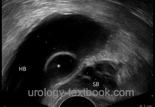
Ureteroceles present as thin-walled cysts in the bladder [fig. ureterocele in TRUS]. For imaging, a medium-filled bladder is preferable. A ureterocele might cause urinary obstruction of the contralateral kidney. Ureteral duplication is often present, and a stenotic ostium of the ureterocele may cause hydronephrosis of the upper pole system.
 |
Intravenous urography:
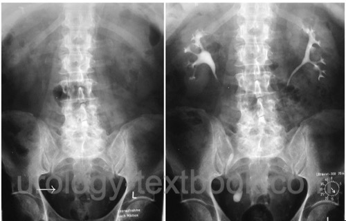
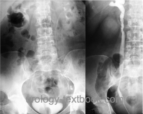
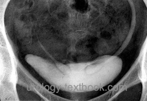
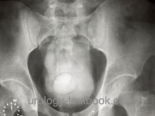
The IVP often shows a poor visualization of the upper renal portion due to poor function; however, the upper part can contrast in late KUB. Hints for a non-contrasting upper pole are reduced chalices and a greater distance between the kidney system and the spine. The lower pole ureter is serpentine (around the upper pole ureter). Sometimes the ureterocele can be visualized in the bladder [fig. ureterocele causes filling defect]. The cystic dilatation of an uncomplicated intravesical ureterocele is called a cobra head sign.
 |
 |
 |
Voiding cystourethrography:
The filling defect by the ureterocele shows size and location. Imaging the ureterocele is best during early filling; the ureterocele may collapse in a full bladder and present as a bladder diverticulum. Filling the bladder to full capacity is necessary to demonstrate reflux into the lower pole ureter, which is seen in duplex kidneys in up to 50%.
Cystoscopy:
See fig. ureterocele in cystoscopy and ureterocele with a stone for findings in cystoscopy.
Retrograde pyelography:
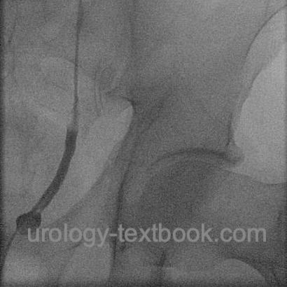
Before invasive treatment, see fig. retrograde pyelography before ureterocele incision and ureterocele with a stone.
Renal scintigraphy:
Renal scintigraphy evaluates renal function or the significance of hydronephrosis. If a duplex system is present, the renal function of the upper and lower pole must be analyzed separately.
Treatment of Ureterocele
A ureterocele without symptoms does not require therapy. The aim is to treat ureterocele with accompanying problems (prevent vesicoureteral reflux and UTI, relieve obstruction) with a single intervention, if possible.
Transurethral Ureterocele Incision:
Transurethral incision of the ureterocele is a treatment option with low morbidity [fig. transurethral incision of an intravesical ureterocele]. It is often curative for obstructive intravesical ureterocele with a single system or with a double system without severe reflux in the lower pole ureter. Extravesical ureteroceles are less likely to be healed by incision, never the less ureterocele incision is often performed as the first treatment attempt. If obstruction or severe postoperative reflux is a problem, ureteropyelostomy or ureteral reimplantation is necessary.
 |
 |
 |
Ureteropyelostomy:
Treatment option for an obstructive ureterocele (relapse after transurethral incision) of a duplex kidney without relevant reflux in the lower pole ureter. The upper pole ureter is anastomosed to the lower pole ureter (end-to-side). Surgery is possible with a flank incision or laparoscopically. The ureterocele should decompress spontaneously and thus reduce the reflux in the lower pole ureter. If postoperative vesicoureteral reflux is problematic, ureterocele resection and ureterocystoneostomy of the lower pole ureter are necessary.
Heminephrectomy for duplex kidneys with ureterocele:
Heminephrectomy is indicated for ureterocele and double system with poor upper pole function and without relevant reflux in the lower pole ureter. Surgery is possible with a flank incision or laparoscopically. The ureterocele should decompress spontaneously and thus reduce the reflux in the lower part. If postoperative vesicoureteral reflux is problematic, ureterocele resection and ureterocystoneostomy of the lower pole ureter are necessary.
Simultaneous reconstruction of the upper and lower urinary tract:
Simultaneous upper and lower urinary tract reconstruction may be necessary for ectopic ureterocele with severe reflux or obstruction of the contralateral kidney. Heminephrectomy or ureteropyelostomy is done via a flank incision, excision of the ureterocele, and ureterocystoneostomy is performed via a separate incision for surgical access to the urinary bladder.
| Ectopic ureter | Index | Vesicoureteral reflux |
Index: 1–9 A B C D E F G H I J K L M N O P Q R S T U V W X Y Z
References
EAU guidelines: Paediatric Urology
Shokeir und Nijman 2002 SHOKEIR, A. A. ; NIJMAN,
R. J.:
Ureterocele: an ongoing challenge in infancy and childhood.
In: BJU Int
90 (2002), Nr. 8, S. 777–83
 Deutsche Version: Diagnose und Therapie der Ureterozele
Urology-Textbook.com – Choose the Ad-Free, Professional Resource
This website is designed for physicians and medical professionals. It presents diseases of the genital organs through detailed text and images. Some content may not be suitable for children or sensitive readers. Many illustrations are available exclusively to Steady members. Are you a physician and interested in supporting this project? Join Steady to unlock full access to all images and enjoy an ad-free experience. Try it free for 7 days—no obligation.
New release: The first edition of the Urology Textbook as an e-book—ideal for offline reading and quick reference. With over 1300 pages and hundreds of illustrations, it’s the perfect companion for residents and medical students. After your 7-day trial has ended, you will receive a download link for your exclusive e-book.