You are here: Urology Textbook > Ureters > Duplicated ureter
Duplicated Ureter or Duplex Kidney: Symptoms and Diagnosis
Definition of Duplex Kidney and Ureteral Duplication
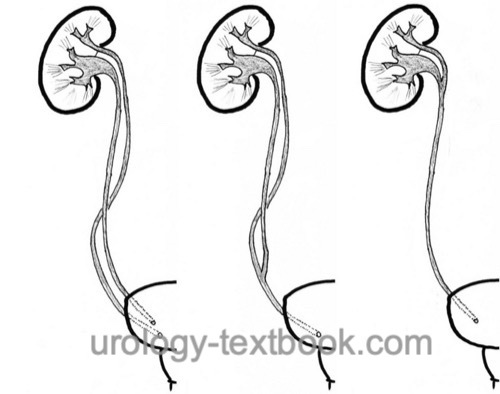
The duplex kidney is an anatomic variant of the ureter and pyelocalyceal system with a large variability [variants of the duplex kidney]:
Complete ureteral duplication:
Duplex kidney with complete duplicated ureters, renal pelvis and two orifices of the ureters.
Ureter fissus or bifid ureter:
Duplex kidney with incomplete ureteral duplication and a common orifice of the ureter.
Bifid renal pelvis:
Duplex kidney with duplication of the renal pelvis and a common ureter.
 |
Inverted Y ureteral duplication:
Two separate ureteral buds merge on the way to the metanephric blastema, resulting in a common renal unit. Very rare.
Ureteral triplication:
Three independent ureteral orifices with three complete ureters or incomplete ureteral triplication with two orifices of the ureters (see ureter fissus).
Epidemiology of the Duplex Kidney
Prevalence 1:150 (0.7%)
Etiology of the Duplex Kidney
Two ureteric buds:
Two ureteric buds on the mesonephric duct (Wolffian duct) result in two separate ureters and a duplex kidney in a common renal capsula. The ureteric buds rotate by 180 degrees while being incorporated into the urogenital sinus: the latero-cranial orifice drains the lower pole of the kidney, and the medio-caudal orifice drains the upper pole of the kidney. This relation is called Weigert-Meyer rule.
Division of the ureteric bud:
Incomplete ureteral duplications (bifid ureter, bifid pelvis) are caused by a division of a single ureteric bud on its way to the metanephric blastema.
Pyelocalyceal system of the duplex kidney:
Compared to a normal kidney with 8–9 calyces, the duplex kidney consists of 11–12 calyces. The upper pole system is smaller, with an average of 3–4 calyces.
Associated malformations:
Vesicoureteral reflux to the lower pole system (40%), ureteropelvic junction obstruction of the lower pole system, ectopic ureteral orifice of the upper pole system (with or without obstruction), ureterocele of the upper pole ureter, hypoplastic and dysplastic renal parenchyma most common of the upper pole system.
Pathophysiology:
Duplication of the ureter is associated with vesicoureteral reflux or hydronephrosis, leading to a susceptibility to urinary tract infections or nephrolithiasis.
Signs and Symptoms of a Duplex Kidney
Associated malformations may lead to urinary tract infections, nephrolithiasis, fever, or abdominal tumor. Without malformations, a duplex kidney usually presents without symptoms.
Diagnostic Workup
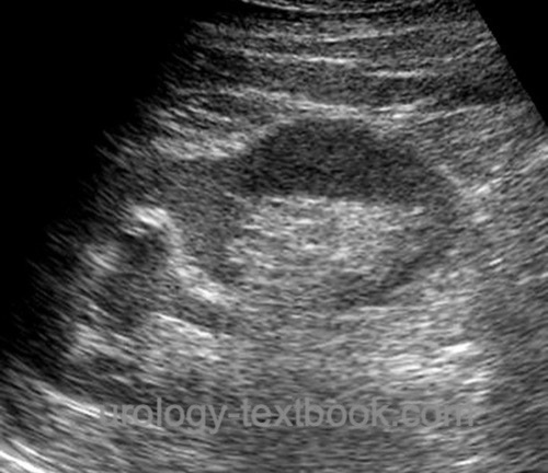
Sonography:
Without associated malformations, a normal finding in renal ultrasound is seen. A duplex kidney may be recognized if renal parenchyma separates two renal pelves (fig. US duplex kidney). It is easier to detect a duplex kidney with ureteroceles or hydronephrosis; most often, the upper pole system is affected (fig. ultrasound of a duplex kidney with hydronephrosis of the upper pole system).
 |
 |
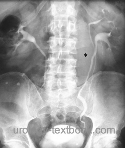
Intravenous urography:
A duplex kidney is often an incidental finding during the diagnostic workup for other diseases [fig. duplex kidney in IVU]. Due to poor renal function, urography often lacks to contrast the upper part of the duplex kidney. Hints for a non-contrasting upper portion are obtained from the small number of calyces shown, inferior-lateral displacement of the lower pole (dropping lily sign) and the greater distance of the renal system to the spine. Sometimes, a ureterocele is delineated in the bladder.
 |
 |
Computed Tomography or Magnetic Resonance Tomography:
The diagnosis and accompanying malformations are reliably detected with CT or MRI, which are increasingly replacing IVP in diagnostics [fig. Ureteral duplication in CT].
 |
Voiding cystourethrogram:
Voiding cystourethrogram is indicated for recurrent urinary tract infections or dilatation of the pyelocalyceal system. A possible ureterocele is detectable in the early filling phase.
Endoscopy:
Cystoscopy, retrograde pyelography, and possibly ureteroscopy depending on complaints and other findings.
Renal scintigraphy:
Renal scintigraphy enables the separate determination of the renal function of the upper and lower part of the duplex kidney for further treatment planning.
Treatment of the Duplex Kidney
Duplex kidney or ureteral duplication is a normal variant and, thus, not subject to treatment. The accompanying malformations and symptoms guide the therapy (vesicoureteral reflux, ureterocele, ectopic ureter, recurrent urinary tract infections, nephrolithiasis, or urinary tract obstruction).
| UPJ obstruction | Index | Ureteral diseases |
Index: 1–9 A B C D E F G H I J K L M N O P Q R S T U V W X Y Z
References
EAU guidelines: Paediatric Urology
 Deutsche Version: Ureteroduplikatur
Urology-Textbook.com – Choose the Ad-Free, Professional Resource
This website is designed for physicians and medical professionals. It presents diseases of the genital organs through detailed text and images. Some content may not be suitable for children or sensitive readers. Many illustrations are available exclusively to Steady members. Are you a physician and interested in supporting this project? Join Steady to unlock full access to all images and enjoy an ad-free experience. Try it free for 7 days—no obligation.
New release: The first edition of the Urology Textbook as an e-book—ideal for offline reading and quick reference. With over 1300 pages and hundreds of illustrations, it’s the perfect companion for residents and medical students. After your 7-day trial has ended, you will receive a download link for your exclusive e-book.