You are here: Urology Textbook > Kidneys > Renal sarcoma
Diagnosis and Treatment of Renal Sarcoma
Renal sarcoma is a rare malignant tumor that arises from mesenchymal cells of the kidney.
Epidemiology
Renal sarcomas account for less than 1 % of all malignant renal tumors. The incidence is approximately 0.5 cases per 1 million inhabitants per year. They occur predominantly in middle-aged and older adults.
Pathology of Renal Sarcomas
Macroscopy:
Renal sarcomas typically appear as large, heterogeneous soft tissue masses with infiltrative growth. Calcifications or areas of fat may be present. Despite an apparently intact pseudocapsule, microscopic infiltration beyond the tumor margin is common and can contribute to local recurrence after incomplete resection.
Histology:
Leiomyosarcoma is the most common type, followed by other soft tissue sarcomas such as liposarcoma, angiosarcoma, undifferentiated pleomorphic sarcoma, rhabdomyosarcoma, and osteosarcoma. Distinguishing a primary renal sarcoma pathologically from sarcomatoid renal cell carcinoma is important and can be difficult.
Metastasis:
Spread is predominantly hematogenous, most commonly to the lungs, and also to the bones and liver. Lymph node metastases are possible but are not typical.
Signs and Symptoms of Renal Sarcomas
Many renal sarcomas are large at diagnosis. Signs of advanced disease include flank pain, a palpable flank mass, and hematuria.
Diagnostic Workup
Abdominal CT or MRI:
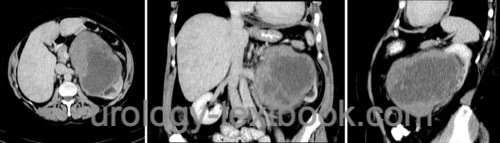
A large, heterogeneous soft tissue mass originating from the kidney [fig. renal sarcoma]. Features that suggest sarcoma include large tumor size, infiltration across anatomical boundaries, and the absence of the typical imaging features of renal cell carcinoma. Extensive calcifications are typical of osteosarcoma. For more information about MRI, see magnetic resonance imaging.
Chest CT:
For preoperative staging.
Tumor biopsy:
Indicated for an atypical mass when the result will guide planning of neoadjuvant therapy.
 |
Differential Diagnosis
Retroperitoneal sarcoma; sarcomatoid renal cell carcinoma; angiomyolipoma with little fat; urothelial carcinoma of the renal pelvis; and other rare renal neoplasms.
Therapy of Renal Sarcomas
Radical nephrectomy:
The goal is complete resection with negative margins, using radical nephrectomy and, when necessary, en bloc resection of infiltrated adjacent organs. Lymphadenectomy is performed when lymph nodes are suspicious clinically or intraoperatively.
Radiotherapy and chemotherapy:
Neoadjuvant and adjuvant treatment strategies should be considered, particularly for large, high-grade, locally advanced, or borderline resectable tumors.
Systemic therapy:
For metastatic disease, systemic treatment depends on the sarcoma subtype. Anthracycline-based chemotherapy remains the most important first-line treatment for many soft tissue sarcomas. Tyrosine kinase inhibitors such as pazopanib may be used after prior treatment for selected non-adipocytic soft tissue sarcomas. Immune checkpoint inhibitors have clinically relevant activity in certain sarcoma subtypes, but evidence for primary renal sarcomas is limited.
Prognosis
Renal sarcoma has a poor prognosis. Important prognostic factors include tumor grade, stage, metastatic disease, and resection with negative margins. In an older series, overall survival at one, three, and five years was 86 %, 41 %, and 15 %, respectively (Wang et al., 2011). Median survival was 28 months; median survival in patients with metastases or after recurrence was 8–10 months.
| Wilms tumor | Index | Renal lymphoma |
Index: 1–9 A B C D E F G H I J K L M N O P Q R S T U V W X Y Z
References
J. Uhlig et al., “Epidemiology, treatment and outcomes of primary renal sarcomas in adult patients,” Sci Rep, vol. 14, no. 1, p. 10038, 2024, doi: 10.1038/s41598-024-60174-8.
Wang, X.; Xu, R.; Yan, L.; Zhuang, J.; Wei, B.; Kang,
D.; Wu, K.; Xie, L.; Xie, Z.; Wei, Q.; Li, X.; Zeng, H. & Lu, Y.
Adult
renal sarcoma: clinical features and survival in a series of patients
treated at a high-volume institution.
Urology 2011, 77, 836-841.
 Deutsche Version: Nierensarkom
Urology-Textbook.com – Choose the Ad-Free, Professional Resource
This website is designed for physicians and medical professionals. It presents diseases of the genital organs through detailed text and images. Some content may not be suitable for children or sensitive readers. Many illustrations are available exclusively to Steady members. Are you a physician and interested in supporting this project? Join Steady to unlock full access to all images and enjoy an ad-free experience. Try it free for 7 days—no obligation.
New release: The first edition of the Urology Textbook as an e-book—ideal for offline reading and quick reference. With over 1300 pages and hundreds of illustrations, it’s the perfect companion for residents and medical students. After your 7-day trial has ended, you will receive a download link for your exclusive e-book.