You are here: Urology Textbook > Ureters > Ectopic ureter
Ectopic Ureter: Diagnosis and Treatment
Definition of Ectopic Ureter
An ectopic ureter is a ureter with an abnormally located ureteral orifice. Instead of draining into the bladder, the ureteral orifice is located in the urethra, vagina, or mesonephric duct structures (ductus deferens or seminal vesicles). EAU guidelines: Paediatric Urology.
Epidemiology
1:2000, more girls than boys.
Etiology
See etiology of ureteral duplication.
Pathology of Ectopic Ureter
80% of ectopic ureters are associated with a ureteral duplication, especially in girls. In boys, the ectopic ureter may also drain a single renal system. In girls, the ectopic ureter drains into the urethra (35%), vulval vestibule (34%), vagina (25%), or uterus (5%). In boys, the ectopic ureter drains into the prostatic urethra (47%), seminal vesicles (33%), prostatic utricle (10%), or vas deferens (10%).
The further the distance of the ureteral opening from its normal position, the higher the likelihood of renal malformation (renal dysplasia, renal hypoplasia) and dysfunction. If the ectopic ureter is associated with a duplex system, the ectopic ureter drains the upper pole of the kidney.
Signs and Symptoms of an Ectopic Ureter
Recurrent urinary tract infections, flank pain, fever, and arterial hypertension. Additional symptoms depend on the gender and location of the ureteral orifice:
- Girls: urinary incontinence (day and night, but sometimes also intermittent), vaginal discharge.
- Boys: LUTS, epididymitis.
Diagnosis of Ureteral Ectopia
Pelvic examination:
Pelvic examination is indicated in girls with urinary incontinence; sometimes, the orifice of the ureter can be identified in the vagina or vulval vestibule.
| Do you want to see the illustration? Please support this website with a Steady membership. In return, you will get access to all images and eliminate the advertisements. Please note: some medical illustrations in urology can be disturbing, shocking, or disgusting for non-specialists. Click here for more information. |
Cystoscopy and retrograde pyelography:
Cystoscopy and retrograde pyelography to search for ureteral orifices.
Ultrasound imaging:
Ureteral ectopia may cause hydronephrosis. If a duplex kidney is present, urinary obstruction of the upper kidney portion may be visible. A dilated ureter may be detectable behind the bladder.
Intravenous Urography:
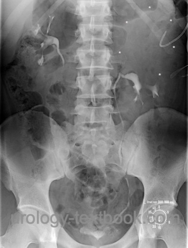
Intravenous urography is increasingly replaced by MR urography, or, in adults, by CT Urography. Due to poor renal function, urography often lacks to contrast the upper part of the duplex kidney. Hints for a non-contrasting upper portion are obtained from the small number of calyces shown, inferior-lateral displacement of the lower pole (dropping lily sign) and the greater distance of the renal system to the spine. Late images after 1–3 hours may show contrast in the upper renal portion.
 |
Voiding Cystourethrography:
VCUG may detect vesicoureteral reflux into the lower renal pole if a duplex kidney is present.
Renal scintigraphy:
Renal scintigraphy is indicated to determine the renal function on the side of the ectopic ureter. If a duplex system is present, the renal function of the upper and lower pole must be analyzed separately.
MRI Urography or CT:
MRI urography is the most accurate imaging tool and is indicated for imaging in children, especially if unclear findings in previous investigations are present and an ectopic ureter is suspected. CT is an imaging alternative in adults that is more sensitive than intravenous urography.
Treatment of Ectopic Ureter
In principle, treatment is only necessary for patients with symptoms, relevant vesicoureteral reflux, or significant obstruction.
Ectopic ureter with sufficient renal function:
- Ureteropyelostomy or ureteroureterostomy for a duplex kidney, usually the ectopic ureter of the upper pole is anastomosed with the lower pole ureter (end-to-side). If significant reflux into the ectopic ureter is present, the resection of the distal ectopic ureter is necessary.
- Common sheath ureteroneocystostomy (UCN) for a duplex kidney with closely spaced ureteral orifices.
- Ureteroneocystostomy (UCN), if the ureter drains a single renal system.
Ectopic ureter with a nonfunctioning kidney:
Standard treatment is heminephrectomy for a double system and nephrectomy for a single system; surgery is possible with a laparoscopic technique. In the case of reflux in the ectopic ureter, an additional distal ureterectomy is necessary. In the case of reflux in the lower part of a duplex kidney, ureteral reimplantation may be necessary.
Heminephrectomy is a complex operation with a small risk of organ loss due to bleeding or urinoma. Alternatively, although the partial function of the upper portion is poor, the ectopic ureter can be anastomosed (ureteroureterostomy or ureteropyelostomy) to the lower pole ureter. The procedure has fewer complications and the prognosis regarding long-term complications (UTI, hypertension) is equal (Kawal et al., 2019.
| Duplex kidney | Index | Ureterocele |
Index: 1–9 A B C D E F G H I J K L M N O P Q R S T U V W X Y Z
References
EAU guidelines: Paediatric Urology
T. Kawal et al., “Ipsilateral ureteroureterostomy: does function of the obstructed moiety matter?,” J Pediatric Urology, vol. 15, no. 1, p. 50, 2019.
 Deutsche Version: Ureterektopie
Urology-Textbook.com – Choose the Ad-Free, Professional Resource
This website is designed for physicians and medical professionals. It presents diseases of the genital organs through detailed text and images. Some content may not be suitable for children or sensitive readers. Many illustrations are available exclusively to Steady members. Are you a physician and interested in supporting this project? Join Steady to unlock full access to all images and enjoy an ad-free experience. Try it free for 7 days—no obligation.
New release: The first edition of the Urology Textbook as an e-book—ideal for offline reading and quick reference. With over 1300 pages and hundreds of illustrations, it’s the perfect companion for residents and medical students. After your 7-day trial has ended, you will receive a download link for your exclusive e-book.