You are here: Urology Textbook > Kidneys > Simple renal ectopia
Simple Renal Ectopia
Definition:
Simple renal ectopia is an abnormal position of the kidney outside the renal fossa in a cranial or caudal direction. It is also termed renal dystopia.
Possible Abnormal Locations of the Kidney:
From cranial to caudal.
Thoracic Kidney:
Location above the diaphragm in the posterior mediastinum. The kidney is separated from the pleural cavity by a thin membrane (thinned diaphragm).
Lumbar Position:
In the iliac fossa.
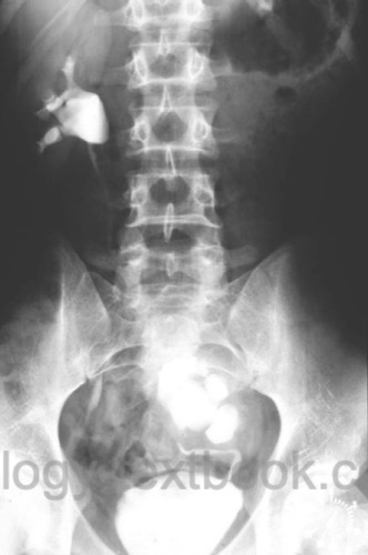
Pelvic Kidney:
Location of the kidney below the aortic bifurcation or in the true pelvis [Fig. left-sided pelvic kidney].
Epidemiology:
1:500 to 1:1200, slightly more common on the left side.
Etiology:
Renal ectopia results from impaired normal embryonic renal ascent. Renal rotation is frequently also disturbed, so that the renal pelvis lies anteriorly rather than medially. Variable, often multiple vascular supply from the distal aorta or iliac vessels is also typical. The cause is usually multifactorial; in addition to disorders of renal ascent, genetic factors and syndromic associations are discussed. The malformation may occur in isolation or together with other urogenital and extrarenal anomalies.
Signs and Symptoms:
Many affected individuals are asymptomatic, and the diagnosis is often made incidentally.
Urological Complications:
Hydronephrosis, often in the setting of ureteropelvic junction obstruction. Urinary tract infections, vesicoureteral reflux, and nephrolithiasis. Arterial hypertension is rare.
Associated Malformations:
Uterine and vaginal malformations are more common in women; cryptorchidism or urethral malformations may occur in men. Extrarenal anomalies may also be associated.
Diagnosis:
The diagnosis is primarily established by ultrasonography. Additional cross-sectional imaging is useful in the presence of unclear anatomy, hydronephrosis, before interventional or surgical treatment, or when relevant vascular variants are suspected.
Therapy:
Treatment is required only for symptoms or complications. In asymptomatic patients with urinary tract dilatation, imaging follow-up is appropriate.
| Supernumerary kidney | Index | Crossed renal ectopia |
Index: 1–9 A B C D E F G H I J K L M N O P Q R S T U V W X Y Z
References
 Deutsche Version: Einfache Nierenektopie
Urology-Textbook.com – Choose the Ad-Free, Professional Resource
This website is designed for physicians and medical professionals. It presents diseases of the genital organs through detailed text and images. Some content may not be suitable for children or sensitive readers. Many illustrations are available exclusively to Steady members. Are you a physician and interested in supporting this project? Join Steady to unlock full access to all images and enjoy an ad-free experience. Try it free for 7 days—no obligation.
New release: The first edition of the Urology Textbook as an e-book—ideal for offline reading and quick reference. With over 1300 pages and hundreds of illustrations, it’s the perfect companion for residents and medical students. After your 7-day trial has ended, you will receive a download link for your exclusive e-book.