You are here: Urology Textbook > Kidneys > Crossed renal ectopia
Crossed Renal Ectopia
Definition of Crossed Renal Ectopia
Crossed renal ectopia is the location of the kidney on the contralateral side in relation to the ureteral orifice location at the bladder. Often, the crossed kidney is fused with the uncrossed kidney [fig. examples of crossed renal ectopia].
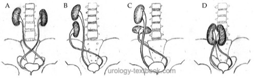 |
Depending on the form and fusion, different forms of crossed renal ectopia are possible (McDonald and McClellan, 1957):
Unilaterally fused kidneys with inferior ectopia:
Most common form of crossed renal ectopia. Both kidneys are in a longitudinal position; the crossed kidney's upper pole fuses to the uncrossed kidney's lower pole. The renal pelvises are directed anteriorly [fig unilateral fused kidneys with inferior ectopia].
S-shaped kidney:
Second most common form of crossed renal ectopia. Both kidneys are in a longitudinal position. The crossed kidney is inferior to the uncrossed kidney, and the renal pelvis is directed laterally. The uncrossed kidney is in a normal position.
L-shaped kidney:
Transversal position of the crossed kidney, the uncrossed kidney lies in a normal position with the lower pole in contact with the crossed kidney.
Disc kidney:
Crossed and uncrossed kidneys are fused with their medial borders; the lateral aspects are normal and create a disc shape. The renal pelvises point anteriorly.
Bilaterally crossed renal ectopia:
Both kidneys are crossed without fusion.
Solitary crossed renal ectopia:
The uncrossed kidney is missing.
 |
Epidemiology:
1:1000 to 1:2000. The most common form is unilaterally fused kidneys with inferior ectopia, slightly more often the crossing from left to right.
Etiology of Crossed Renal Ectopia
The etiology is not fully understood; proposed mechanisms include misdirected growth of the ureteric bud, disturbances of renal ascent, vascular barriers, and other mechanical factors.
Signs and Symptoms of Crossed Renal Ectopia
Usually asymptomatic; the diagnosis is often made prenatally or incidentally. Symptoms typically arise only when complications develop (hydronephrosis, UTI, VUR, or nephrolithiasis) or when associated malformations are present.
Diagnosis
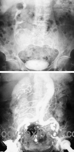
- Ultrasound: Key findings are an empty renal fossa on one side and two kidneys or one large misshapen kidney on the contralateral side.
- CT or MRI: Additional cross-sectional imaging is useful when the anatomy is unclear, in the presence of hydronephrosis, or before interventional or surgical treatment.
Treatment of Crossed Renal Ectopia
Treatment is only necessary in the presence of symptoms or complications. In asymptomatic patients with urinary tract ectasia, imaging follow-up is advisable.
| Simple renal ectopia | Index | Horseshoe kidney |
Index: 1–9 A B C D E F G H I J K L M N O P Q R S T U V W X Y Z
References
McDonald und McClellan 1957 MCDONALD, J. H. ; MCCLELLAN, D. S.: Crossed renal ectopia.In: Am J Surg
93 (1957), Nr. 6, S. 995–1002
 Deutsche Version: gekreuzte Nierenektopie
Urology-Textbook.com – Choose the Ad-Free, Professional Resource
This website is designed for physicians and medical professionals. It presents diseases of the genital organs through detailed text and images. Some content may not be suitable for children or sensitive readers. Many illustrations are available exclusively to Steady members. Are you a physician and interested in supporting this project? Join Steady to unlock full access to all images and enjoy an ad-free experience. Try it free for 7 days—no obligation.
New release: The first edition of the Urology Textbook as an e-book—ideal for offline reading and quick reference. With over 1300 pages and hundreds of illustrations, it’s the perfect companion for residents and medical students. After your 7-day trial has ended, you will receive a download link for your exclusive e-book.