You are here: Urology Textbook > Kidneys > Nephrolithiasis
Nephrolithiasis: Epidemiology and Etiology
- Nephrolithiasis: Etiology and Epidemiology
- Diagnosis of Nephrolithiasis and Renal Colic
- Treatment of Kidney and Ureteral Stones
Definition
Nephrolithiasis is the formation of calculi in the renal pelvic calyceal system; stones may also migrate into the urinary tract (urolithiasis). EAU guidelines: Urolithiasis. Nephrolithiasis should be distinguished from bladder stones, which primarily arise in the bladder; see bladder stones.
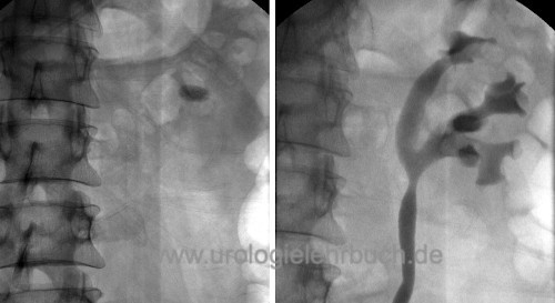 |
Epidemiology of Nephrolithiasis
Diseases of affluence:
There is an increased risk of nephrolithiasis in adults in societies without food shortages and with reduced physical activity (risk factor: metabolic syndrome). In children, urinary stones are more common worldwide in regions with dehydration, infections, malnutrition, or endemic bladder stones.
Risk of disease:
The incidence of nephrolithiasis is approximately 0.5 % in Europe and the USA (500 per 100,000 persons per year). The lifelong risk for nephrolithiasis is 10–15 %. The recurrence risk within 10 years is 50 % (Moe et al., 2006). More than half of stone formers have only one recurrence; 10–20 % experience three or more recurrences during their lifetime. The male-to-female ratio is 3:1, and familial clustering is common.
Etiology and Pathogenesis of Nephrolithiasis
Pathogenesis
Matrix:
Stone crystals are deposited secondarily on an organic matrix to form calculi. The matrix accounts for 2–10 % of a mature stone.
Crystallization:
Five mechanisms lead to the formation of stones:
- Supersaturation
- Nucleation
- Crystal growth
- Aggregation of crystals
- Deficiency of inhibitors of nucleation, crystallization, and aggregation promotes stone formation; these inhibitors include: pyrophosphate, citrate, magnesium, glycosaminoglycans, heparin, and chondroitin sulfate.
Calcium Nephrolithiasis
Calcium-containing stones are the most common type of urinary stone and are usually caused by hypercalciuria. Hypercalciuria is variably defined as urinary calcium excretion of > 5–7.5 mmol (200–250 mg)/24 h. Fifty percent of serum calcium (almost 100 % of ionized calcium) is filtered by the glomeruli; most is reabsorbed by the tubules, and less than 2% is excreted.
Stone types:
Calcium-containing stones are:
- Weddellite stones (calcium oxalate dihydrate)
- Whewellite stones (calcium oxalate monohydrate)
- Hydroxyapatite stones (calcium phosphate)
- Brushite stones (calcium hydrogen phosphate)
Absorptive hypercalciuria:
The regular food intake contains 500–1000 mg of calcium; typically, 150–200 mg are absorbed in the small intestine and later excreted via the urine. With increased calcium absorption in the small intestine, the calcium concentration in the blood increases, which leads to a reduced parathyroid hormone concentration. The reduced parathyroid hormone level decreases tubular calcium reabsorption and thereby causes hypercalciuria.
Resorptive (hormonal) hypercalciuria:
Primary hyperparathyroidism leads to hypercalcemia due to increased calcium resorption from the bone and absorption from the intestine: hypercalciuria, nephrocalcinosis, and recurrent nephrolithiasis develop.
Renal hypercalciuria:
Tubular defects can cause hypercalciuria. Increased renal calcium excretion may lead secondarily to increased parathyroid hormone levels, increased vitamin D activity, mobilization of calcium from bone, and increased intestinal calcium absorption.
Monogenic causes of hypercalciuria:
See the table Monogenic causes of hypercalciuria.
Hyperuricosuria:
Hyperuricosuria promotes the formation of uric acid stones (see below) and increases the risk for calcium stones. Hyperuricosuria reduces the efficiency of inhibitors of lithogenesis, and typically calcium oxalate stones are formed.
Hyperoxaluria:
Increased urinary oxalate excretion (> 0.5 mmol/24 h urine) leads to calcium oxalate stones.
Secondary hyperoxaluria:
Increased enteral oxalate absorption arises from chronic diarrheal states or steatorrhea, such as in ileum or pancreatic diseases. The fats in the intestine consume the calcium in the intestine during saponification processes, which normally binds the oxalate and prevents absorption. Other causes are dietary factors such as foods rich in oxalate (spinach, rhubarb, chard) or vitamin C supplements.
Primary hyperoxaluria:
Due to enzyme defects in the liver, there is a massive increase in oxalic acid, which is deposited in various tissues. Oxaluria is typically over 1 mmol/24 h urine. Depending on the genetic defect, the disease can lead to end-stage renal failure.
Hypocitraturia:
Citrate is an inhibitor of lithogenesis by complexing with calcium and hindering calcium salt crystallization. Hypocitraturia is found in 20–60 % of the calcium stone carriers alone or in combination with other metabolic disorders as the cause of the stone formation.
Reduced citrate excretion usually results from an increased acid load: distal renal tubular acidosis (see below), hypokalemia, chronic diarrhea, hypomagnesemia, fasting, excessive meat consumption, androgen abuse and excessive physical training (bodybuilder stones). The bacteria in urinary tract infections can also consume citrate.
Low urine pH:
Persistently acidic urine with a pH below 6 is a risk factor for calcium-containing kidney stones. This pattern is associated with metabolic syndrome, insulin resistance in type 2 diabetes mellitus, and high animal protein intake.
Renal tubular acidosis (RTA):
Renal tubular acidosis is a group of tubular disorders of proton excretion or bicarbonate absorption with the formation of metabolic acidosis.
Type 1 (distal) RTA:
Type 1 RTA is a disturbed proton secretion of the distal nephron (a defect in type A intercalated cells of the collecting ducts) with the development of hypokalemic, hyperchloremic metabolic acidosis, nephrolithiasis, nephrocalcinosis, and increased urine pH. Distal RTA is a heterogeneous disorder with different mutations, enzyme defects, and phenotypes. Typically, the daily profile of the urine pH always shows values above 5.8, and calcium phosphate stones are formed. Even after exposure to acid (ammonium chloride 0.1 g/kg body weight), the urine pH remains above 5.4. Depending on the level of metabolic acidosis, a distinction is made between incomplete (normal bicarbonate concentration in the BGA) or complete (reduced bicarbonate concentration) renal tubular acidosis.
Type 2 (proximal) RTA:
A defect of bicarbonate absorption in the proximal tubule leads to growth retardation in children, hypokalemia, metabolic acidosis, and metabolic bone disease. Citrate excretion is usually normal; nephrolithiasis typically does not develop.
Type 4 (distal) RTA:
Type 4 RTA usually results from chronic kidney disease (e.g., diabetes mellitus) with reduced sodium reabsorption and insufficient proton and potassium excretion. In addition, there is resistance to aldosterone. Nephrolithiasis is not typical.
Other Types of Nephrolithiasis
Infection stones:
Infection stones account for 10 % of urinary stones. They consist of magnesium, ammonium, and phosphate (MAP; struvite), but may also contain calcium phosphate. Urinary tract infections with urea-splitting bacteria (enzyme urease) cause ammonium production and alkaline urine (> 7), which causes MAP crystals to precipitate. The most common urease-producing bacteria in urinary tract infections are Proteus, Klebsiella, Pseudomonas, and Staphylococcus. Infection stones are often large (staghorn calculi), especially if several clinical risk factors are present: urinary malformations, diabetes, neurologic disorders, immobility, spinal cord injury, and indwelling catheters.
Uric acid stones:
Uric acid stones account for 10 % of urinary stones. They are radiolucent on conventional abdominal radiographs but produce a typical acoustic shadow on ultrasound and are visible on computed tomography. Uric acid stones form in the presence of hyperuricosuria and acidic urine (pH below 6). Low urine volume is also a risk factor. Many disorders cause these risk factors; persistently acidic urine is the most important pathophysiologic factor.
Hyperuricosuria:
Hyperuricosuria is defined as daily uric acid excretion above 4 mmol/24 h; hyperuricemia is not always present. Hyperuricosuria is caused by increased purine intake with food, endogenous overproduction due to enzyme defects, myeloproliferative diseases, tumor lysis syndrome, chemotherapy, medication (probenecid), or catabolic situations.
Cystine stones:
Cystine stones account for 1–2 % of urinary stones. Heterozygous (1:200) and homozygous (1:20,000) cystinuria may lead to cystine stone precipitation in acidic urine when urinary cystine excretion exceeds 0.8 mmol/24 h. A rounded, frosted-glass appearance is typical on radiographs. Cystine stones are unsuitable for therapy with ESWL.
Xanthine stones:
Xanthine stones result from a congenital xanthine oxidase defect. Like uric acid stones, they are radiolucent.
2,8-Dihydroxyadenine stones:
Children with congenital adenine phosphoribosyltransferase (APRT) deficiency form 2,8-dihydroxyadenine stones.
Indinavir stones:
Indinavir, an HIV antiretroviral medication, may cause stones that are radiolucent and not reliably detected by CT.
| Kidney transplantation | Index | Renal colic |
Index: 1–9 A B C D E F G H I J K L M N O P Q R S T U V W X Y Z
References
Coe u.a. 2005 COE, F. L. ; EVAN, A. ;
WORCESTER, E.:
Kidney stone disease.
In: J Clin Invest
115 (2005), Nr. 10, S. 2598–608
EAU Guidelines: Urolithiasis [Online]. https://uroweb.org/guidelines/urolithiasis/
Moe 2006 MOE, O. W.:
Kidney stones: pathophysiology and medical management.
In: Lancet
367 (2006), Nr. 9507, S. 333–44
 Deutsche Version: Epidemiologie und Ursachen der Nephrolithiasis
Urology-Textbook.com – Choose the Ad-Free, Professional Resource
This website is designed for physicians and medical professionals. It presents diseases of the genital organs through detailed text and images. Some content may not be suitable for children or sensitive readers. Many illustrations are available exclusively to Steady members. Are you a physician and interested in supporting this project? Join Steady to unlock full access to all images and enjoy an ad-free experience. Try it free for 7 days—no obligation.
New release: The first edition of the Urology Textbook as an e-book—ideal for offline reading and quick reference. With over 1300 pages and hundreds of illustrations, it’s the perfect companion for residents and medical students. After your 7-day trial has ended, you will receive a download link for your exclusive e-book.