You are here: Urology Textbook > Kidneys > Horseshoe kidney
Horseshoe Kidney
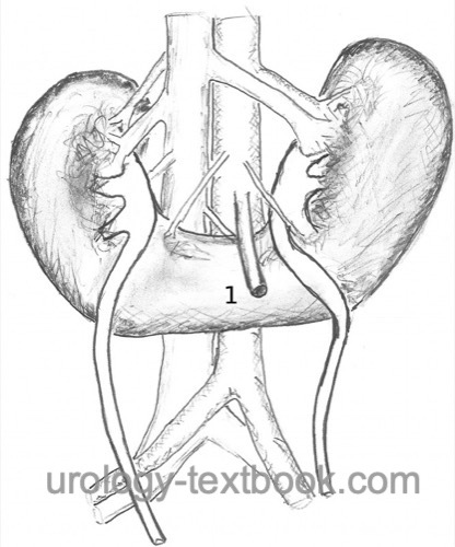
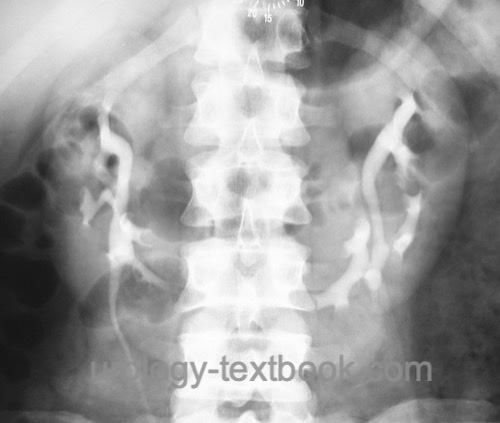
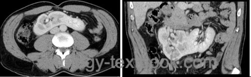
A horseshoe kidney is the most common congenital renal fusion anomaly. The lower poles of both kidneys are fused across the midline [see the following figures]. The fusion, usually consisting of renal parenchyma, is termed the isthmus. Fusion interferes with renal rotation and ascent; therefore, horseshoe kidneys are usually positioned lower than normal and the renal pelves are oriented anteriorly (Stein and Desai, 2007).
Epidemiology:
Incidence 1:700. Male-to-female ratio 2:1.
Etiology of Horseshoe Kidney
The lower poles fuse before renal rotation and ascent; consequently, the renal pelves are oriented anteriorly and the calyceal system posteriorly. The inferior mesenteric artery prevents further renal ascent. The vascular supply from the iliac vessels and distal aorta persists in a horseshoe kidney. The abnormal vascular supply can cause extrinsic ureteropelvic junction obstruction with hydronephrosis.
Signs and Symptoms of Horseshoe Kidney
Many patients remain asymptomatic, and the diagnosis is often made incidentally or prenatally. Otherwise, hydronephrosis is the most common finding, followed by nephrolithiasis, vesicoureteral reflux, or infection.
Risk of associated malformations:
Horseshoe kidney is frequently associated with other malformations, including urogenital anomalies, neural tube defects, congenital heart defects, and musculoskeletal anomalies. Horseshoe kidney occurs more frequently in chromosomal syndromes such as Turner syndrome or trisomy 18.
Oncologic risk:
The risk of Wilms tumor is doubled. The very rare renal carcinoid occurs more frequently in horseshoe kidneys.
Diagnosis
Typical ultrasound findings are low-lying kidneys, fusion of the lower poles by an isthmus anterior to the aorta, and anteriorly oriented renal pelves. Additional cross-sectional imaging (CT or MRI) is useful if the anatomy is unclear, before interventional or surgical treatment, or if complications are suspected.
Treatment of Horseshoe Kidney
Nephrolithiasis:
All stone-treatment techniques can be used: extracorporeal shock wave lithotripsy (ESWL), ureteroscopy, or percutaneous nephrolithotomy (Stein and Desai, 2007). The atypical position and rotational anomaly of the horseshoe kidney must be considered.
Hydronephrosis:
With preserved renal function and clinically relevant ureteropelvic junction obstruction, pyeloplasty is indicated. With severely reduced differential renal function, heminephrectomy is rarely necessary.
Tumors of the horseshoe kidney:
Treatment depends on tumor size, patient age, and suspected histology. During surgical treatment, the atypical position and abnormal vascular supply must be considered. Careful preoperative imaging and planning of the surgical approach are essential.
| Kidney diseases | Index | Kidney malrotations |
Index: 1–9 A B C D E F G H I J K L M N O P Q R S T U V W X Y Z
References
Kölln, C. P.; Boatman, D. L.; Schmidt, J. D. & Flocks, R. H. Horseshoe kidney: a review of 105 patients.J Urol, 1972, 107, 203-204.
Stein, R. J. & Desai, M. M. Management of urolithiasis in the congenitally abnormal kidney (horseshoe and ectopic).
Curr Opin Urol, 2007, 17, 125-131.
 Deutsche Version: Hufeisenniere
Urology-Textbook.com – Choose the Ad-Free, Professional Resource
This website is designed for physicians and medical professionals. It presents diseases of the genital organs through detailed text and images. Some content may not be suitable for children or sensitive readers. Many illustrations are available exclusively to Steady members. Are you a physician and interested in supporting this project? Join Steady to unlock full access to all images and enjoy an ad-free experience. Try it free for 7 days—no obligation.
New release: The first edition of the Urology Textbook as an e-book—ideal for offline reading and quick reference. With over 1300 pages and hundreds of illustrations, it’s the perfect companion for residents and medical students. After your 7-day trial has ended, you will receive a download link for your exclusive e-book.