You are here: Urology Textbook > Anatomy > Kidney > Embryology
Embryology: Development of the Kidney and Ureter
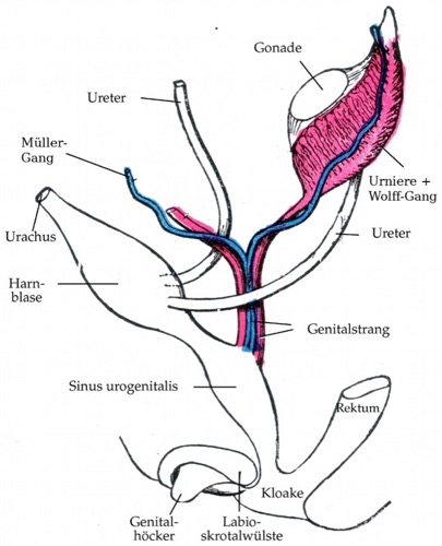
Development of the Pronephros and Mesonephros
The urogenital system develops from the intermediate mesoderm, which lies between the paraxial mesoderm (somites) and the lateral plate mesoderm. The intermediate mesoderm forms the nephrogenic cord in a segmented, bead-like pattern.
Pronephros
The pronephros develops from cervical segments of the nephrogenic cord. In humans, it remains rudimentary and is no longer detectable by the end of the fourth embryonic week. The pronephric duct is incorporated into the mesonephric system and persists as the mesonephric duct (Wolffian duct, ductus mesonephricus).
Mesonephros
The mesonephros develops from subsequent thoracic and upper lumbar segments. Each segment forms multiple mesonephric tubules with renal corpuscles (glomeruli) and a tubular system. The mesonephros is transiently functional and drains urine via the mesonephric (Wolffian) duct. Regression typically begins at approximately week 7.
- The ureteric bud arises from the distal mesonephric (Wolffian) duct and, through reciprocal signaling with the metanephric mesenchyme, initiates formation of the definitive kidney (see below).
- In male development, the mesonephric (Wolffian) duct persists and forms the excurrent duct system, and selected mesonephric tubules contribute to the efferent ductules (see the corresponding section).
- In female development, the mesonephros and mesonephric duct regress, leaving only a few remnants.
 |
Metanephros (Definitive Kidney)
The definitive kidney (metanephros) develops through reciprocal interactions between the ureteric bud and the metanephric mesenchyme. In the fifth embryonic week, the ureteric bud sprouts from the mesonephric (Wolffian) duct near its entry into the cloaca. The ureteric bud gives rise to the collecting system (ureter, renal pelvis, calyces, and collecting ducts), which grows into the metanephric blastema and induces metanephric differentiation.
The cranial end of the ureteric bud (the ampulla) undergoes dichotomous branching and induces proliferation and differentiation of the metanephric mesenchyme. Early branches form the renal pelvis and the calyceal system (typically about 10–25 minor calyces with corresponding papillae). Early splitting of the ureteric bud or the presence of additional buds can produce duplication anomalies (e.g., ureteral duplication and a duplicated renal pelvis). Continued branching forms the collecting duct system. At each papilla, multiple collecting ducts open as papillary ducts (ducts of Bellini) at the papillary tip. Branching continues into the second trimester. Each terminal collecting duct branch ends in a terminal ampulla, where it induces the formation of a nephron primordium. Failure of connection between a developing nephron and the collecting system can contribute to cyst formation.
Nephrogenesis typically ends near the end of gestation (approximately weeks 34–36). By birth, the kidney has established its final nephron endowment, averaging about 1,000,000 renal corpuscles per kidney, although the range is wide. Nephron number correlates with birth weight. A reduced nephron endowment increases the risk of chronic kidney disease and arterial hypertension.
During early development (approximately weeks 6 through 9), the kidneys ascend as the embryo grows. The kidneys also rotate along their long axis so that the renal hilum shifts from an anterior to a medial orientation. Malrotation of the kidney can occur in any direction, and renal malrotation is common if other abnormalities are present. As ascent proceeds, the arterial supply changes from branches of the iliac vessels to direct branches of the abdominal aorta. The definitive renal artery usually arises at the L1–L2 level. This stepwise revascularization commonly results in variations of the renal vasculature.
| Urologic Surgery | Index | Embryology of the testis |
Index: 1–9 A B C D E F G H I J K L M N O P Q R S T U V W X Y Z
References
Benninghoff, A. (1993): Makroskopische Anatomie, Embryologie und Histologie des Menschen. 15. Auflage. München; Wien; Baltimore: Urban & Schwarzenberg.
 Deutsche Version: Embryologie: Entwicklung der Nieren
Urology-Textbook.com – Choose the Ad-Free, Professional Resource
This website is designed for physicians and medical professionals. It presents diseases of the genital organs through detailed text and images. Some content may not be suitable for children or sensitive readers. Many illustrations are available exclusively to Steady members. Are you a physician and interested in supporting this project? Join Steady to unlock full access to all images and enjoy an ad-free experience. Try it free for 7 days—no obligation.
New release: The first edition of the Urology Textbook as an e-book—ideal for offline reading and quick reference. With over 1300 pages and hundreds of illustrations, it’s the perfect companion for residents and medical students. After your 7-day trial has ended, you will receive a download link for your exclusive e-book.