You are here: Urology Textbook > Anatomy > Testis > Embryology
Embryology: Development and Descent of the Testis
Embryology of the Indifferent Gonadal Primordium
Early development is initially sex-nonspecific and proceeds through an indifferent gonadal stage. Beginning around the seventh week of gestation, the indifferent gonad differentiates into a testis or an ovary. Thereafter, gonadal hormones and additional signaling pathways direct sex-specific development of the internal and external genitalia.
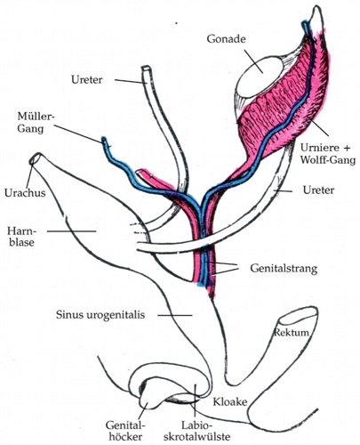 |
The gonadal primordium develops adjacent to the mesonephros. It comprises three major cell populations: cells derived from the coelomic epithelium, cells from the mesenchyme of the mesonephric region, and primordial germ cells. Primordial germ cells become segregated from the somatic cell lineages very early (epiblast stage) in embryogenesis. During subsequent development, they proliferate and migrate via endoderm, yolk sac, hindgut, dorsal mesentery into the gonadal primordium during weeks 5 to 6.
Embryology of the Testis
Testis development requires the presence of the Y chromosome and expression of the SRY gene (sex-determining region of the Y chromosome). SRY encodes a transcription factor (testis-determining factor, TDF) that initiates a downstream gene network driving male sex differentiation (e.g., SOX9, SF1/NR5A1, WT1). Loss of SRY function can result in gonadal dysgenesis with a female or ambiguous phenotype in an individual with a 46,XY karyotype.
Beginning around week 7, testis cords form in the central region of the indifferent gonad as precursors of the seminiferous tubules. Germ cells enter these cords and differentiate into prospermatogonia. The cords lose their connection to the surface epithelium, and a primary tunica albuginea forms. Fetal Leydig cells differentiate from the interstitium (approximately from week 8), and their proliferation enables fetal testosterone production. After birth, the fetal Leydig cell population largely regresses, androgen production declines, and it remains low until puberty.
Sertoli cells in the testis produce anti-Müllerian hormone (AMH), which induces regression of the Müllerian ducts (paramesonephric ducts) and thereby prevents development of the internal female reproductive tract.
Testicular Descent
Early in development, the testis lies retroperitoneally on the posterior abdominal wall and is connected to the mesonephric region by a mesorchium. The caudal gonadal ligament (gubernaculum testis) develops from the caudal peritoneal fold and associated connective tissue and extends toward the labioscrotal swelling. As the body grows and testicular descent proceeds in two phases, the testis shifts caudally while maintaining its vascular supply via the testicular artery from the aorta. The inguinoscrotal phase of descent typically begins around the seventh month of gestation. In the scrotum, the testis becomes surrounded by coverings derived from the layers of the abdominal wall. The peritoneum evaginates into the scrotum as the processus vaginalis. The distal portion persists as the tunica vaginalis (the serous cavity around the testis), whereas the proximal portion normally obliterates.
Embryology of the Excurrent Duct System
The Wolffian duct (mesonephric duct), which initially functions as the drainage duct of the mesonephros, empties into the cloaca (and later into structures derived from the urogenital sinus). Under androgen stimulation, the Wolffian duct differentiates into the epididymal duct, vas deferens, and outgrowths that form the seminal vesicles in the distal mesonephric duct region [Fig. male genital primordium]. A cranial blind-ending remnant can persist as the appendix epididymidis. Several persistent mesonephric tubules are stimulated to proliferate and form the efferent ductules (ductuli efferentes) in the head of the epididymis. These later connect the rete testis to the epididymal duct. The vas deferens ultimately joins the duct of the seminal vesicle to form the ejaculatory duct, which opens into the prostatic urethra.
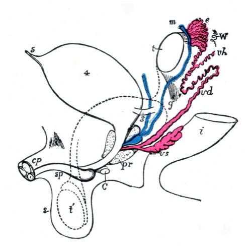 |
Aberrant mesonephric tubules that fail to connect to the rete testis or detach from the Wolffian duct can promote cystic appendages and epididymal cysts. Remnants of mesonephric tubules can also form the paradidymis (organ of Giraldés). This rudimentary structure is most often identified in children and either regresses or may present as a cystic lesion.
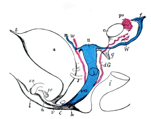
The paired Müllerian duct (paramesonephric duct) is an additional embryonic primordium of the reproductive tract alongside the Wolffian duct and gives rise to the internal female reproductive organs [Fig. female genital primordium]. The Müllerian ducts initially lie lateral to the Wolffian ducts, fuse caudally, and contact the urogenital sinus. Under the influence of AMH produced by Sertoli cells, the Müllerian ducts regress in male embryos. Typical Müllerian remnants include the appendix testis (hydatid of Morgagni) and the prostatic utricle.
 |
| Embryology of the kidney | Index | Embryology lower urinary tract |
Index: 1–9 A B C D E F G H I J K L M N O P Q R S T U V W X Y Z
References
Benninghoff, A. (1993): Makroskopische Anatomie, Embryologie und Histologie des Menschen. 15. Auflage. München; Wien; Baltimore: Urban & Schwarzenberg.
 Deutsche Version: Embryologie: Entwicklung der Hoden
Urology-Textbook.com – Choose the Ad-Free, Professional Resource
This website is designed for physicians and medical professionals. It presents diseases of the genital organs through detailed text and images. Some content may not be suitable for children or sensitive readers. Many illustrations are available exclusively to Steady members. Are you a physician and interested in supporting this project? Join Steady to unlock full access to all images and enjoy an ad-free experience. Try it free for 7 days—no obligation.
New release: The first edition of the Urology Textbook as an e-book—ideal for offline reading and quick reference. With over 1300 pages and hundreds of illustrations, it’s the perfect companion for residents and medical students. After your 7-day trial has ended, you will receive a download link for your exclusive e-book.