You are here: Urology Textbook > Kidneys > Renal angiomyolipoma
Diagnosis and Treatment of Renal Angiomyolipomas
Definition of Angiomyolipoma of the Kidney
Angiomyolipoma of the kidney is a rare benign renal tumor with a high-fat content (Oesterling et al., 1986) (Nelson et al., 2002).
 |
Epidemiology of Renal Angiomyolipoma
Prevalence:
The prevalence in autopsies is 0.3%, with ultrasound screening at 0.1%. Women are much more frequently affected than men. Age peak 50–60 years.
Tuberous sclerosis:
45–80% of patients with tuberous sclerosis have (bilateral) asymptomatic angiomyolipomas. There is an equal distribution between men and women in tuberous sclerosis, with an age peak of 30 years.
Etiology and Pathology of Angiomyolipoma
Etiology:
Angiomyolipomas derive from perivascular epithelioid cells and grow probably hormone-dependent.
Gross Pathology of Angiomyolipoma:
Angiomyolipomas are grey-yellow lesions without a tumor capsule, round to oval. Sometimes, angiomyolipomas show multi-center growth involving lymph nodes without metastatic potential.
Histology of Angiomyolipoma:
Mature fat cells, smooth muscle cells, (atypical) blood vessels, and occasional mitoses.
Signs and Symptoms of Angiomyolipoma of the Kidney
- The vast majority is symptom-free, flank pain is possible.
- Potentially life-threatening hemorrhage due to spontaneous rupture into the retroperitoneum (Wunderlich's syndrome). Pregnancy increases the risk of rupture.
Diagnosis of Renal Angiomyolipoma
Sonography of the Kidney:
Sonography of the kidneys shows an echogenic mass in the kidney caused by the high-fat content. Since all other kidney tumors generate hypoechoic to isoechogenic lesions, it is possible to differentiate angiomyolipoma from other kidney tumors by ultrasound imaging.
 |
 |
 |
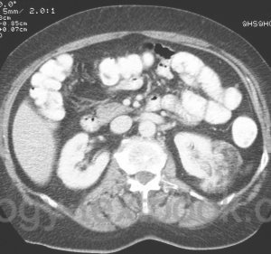
CT-Abdomen:
The fat content typically causes a hypodense mass in the kidney with -20 to -80 HU enabling the differentiation to renal cell carcinoma [fig. CT1 renal angiomyolipoma and CT2 renal angiomyolipoma]. Calcifications are rare in renal angiomyolipoma.
 |
MRI Abdomen:
MRI enables the reliable detection of fat, which is typical for angiomyolipoma, and allows the differentiation to a renal cell carcinoma.
Angiography:
Neovascularization, similar to renal cell carcinoma, is possible. Angiography has no role in the differential diagnosis of renal tumors anymore.
Treatment of Renal Angiomyolipomas
Conservative Treatment of Angiomyolipomas:
Annual imaging controls are possible with angiomyolipomas less than 4 cm and without symptoms. The annual growth rate of sporadic angiomyolipoma is less than 2 mm/year but significantly higher in tuberous sclerosis (Chan et al., 2018).
Partial Nephrectomy:
Partial nephrectomy is indicated in angiomyolipomas of >4 cm, with symptoms or tumors of uncertain dignity.
Selective embolization:
Selective embolization is a possible minimally invasive treatment option. Sometimes, a relapse is possible after embolization. Rarely percutaneous drainage of necrosis is necessary.
Emergency therapy for retroperitoneal hemorrhage:
Depending on the intensity of the hemorrhage, surgical therapy (with partial resection or nephrectomy) or selective arterial embolization of the hemorrhage.
| Renal oncocytoma | Index | Renal diseases |
Index: 1–9 A B C D E F G H I J K L M N O P Q R S T U V W X Y Z
References
Nelson, C. P. & Sanda, M. G.
Contemporary
diagnosis and management of renal angiomyolipoma.
J Urol, 2002,
168, 1315-1325
Oesterling u.a. 1986 OESTERLING, J. E. ;
FISHMAN, E. K. ; GOLDMAN, S. M. ; MARSHALL, F. F.:
The management of renal angiomyolipoma.
In: J Urol
135 (1986), Nr. 6, S. 1121–4
 Deutsche Version: Angiomyolipom der Nieren
Urology-Textbook.com – Choose the Ad-Free, Professional Resource
This website is designed for physicians and medical professionals. It presents diseases of the genital organs through detailed text and images. Some content may not be suitable for children or sensitive readers. Many illustrations are available exclusively to Steady members. Are you a physician and interested in supporting this project? Join Steady to unlock full access to all images and enjoy an ad-free experience. Try it free for 7 days—no obligation.
New release: The first edition of the Urology Textbook as an e-book—ideal for offline reading and quick reference. With over 1300 pages and hundreds of illustrations, it’s the perfect companion for residents and medical students. After your 7-day trial has ended, you will receive a download link for your exclusive e-book.