You are here: Urology Textbook > Anatomy > Kidney > Histology of the Nephron (Glomerulus)
Histology of the kidney (2/7): Nephron and Glomerulus
- Anatomy of the kidney (1/7): Gross anatomy
- Anatomy of the kidney (2/7): Histology of the glomerulus and nephron
- Anatomy of the kidney (3/7): Histology of renal tubules
- Anatomy of the kidney (4/7): Physiology of the glomerular filtration rate
- Anatomy of the kidney (5/7): Physiology of the tubular reabsorption
- Anatomy of the kidney (6/7): Physiology of the renin-angiotensin-aldosterone system
- Anatomy of the kidney (7/7): Physiology of erythropoetin, endothelins and vitamin D
Review literature: (Benninghoff, 1993).
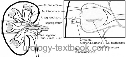
Vascular Architecture of the Kidney
The renal artery branches in five segmental arteries, from which the interlobar arteries arise. The interlobar arteries run between the renal pyramids to the border of the renal cortex and medulla and partition into the arcuate arteries. The arcuate arteries pass along the cortical-medullary border, parallel to the lateral convex border of the kidney. The arcuate arteries divide into the interlobular arteries, which run into the cortex to form the afferent arterioles and supply the glomeruli [fig. renal blood flow]. The arcuate arteries also give branches to the vasa recta (see below)
The blood leaves the glomerulus (first capillary microcirculation) through the efferent arterioles. The efferent arteriolar near the medulla run into the medulla and form the vasa recta to supply the tubules in the renal medulla (second capillary microcirculation). The efferent arterioles near the cortex supply the tubules of the cortex.
The vasa recta run parallel to the tubules of the medulla from the cortex-medulla border to the papilla and back. This results in a countercurrent vascular architecture, which enables the high osmotic gradient in the medulla. The venae rectae drain into the arcuata veins, which drain via the interlobular veins to the renal vein.
 |
The Nephron
Each kidney has about 1 million functional units, the nephrons [schematic figure of the nephron]. The number of nephrons is considerably variable, e.g., the number of nephrons correlates with birth weight. A low number of nephrons carry an increased risk for kidney disease and hypertension.
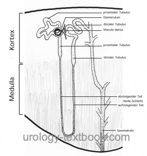 |
A nephron is divided into the following subsections, which are described on the following pages:
- Renal corpuscle (glomerulus)
- Proximal tubule
- Proximal Convoluted Tubule (PCT) or pars convoluta
- Proximal Straight Tubule (PST) or pars recta
- Loop of Henle
- Descending Thin Limb (DTL)
- Ascending Thin Limb (ATL)
- Thick Ascending Limb (TAL)
- Distal tubule
- Thick Ascending Limb (TAL)
- Macula densa
- Distal Convoluted Tubule (DCT)
- Collecting Duct (CD)
Glomerulus (Renal corpuscles)
The kidney consists of about 1 000 000 renal corpuscles, which corresponds to the number of nephrons. The number of nephrons is considerably variable. Depending of the location of the glomerulus, nephrons are classified as cortical nephrons or juxtamedullary nephrons.
The afferent arterioles supply the glomerular capillaries with blood. There is a constant proportion of the blood plasma (primary urine), which is filtrated in the Bowman space of the glomerulus. The primary urine passes from the Bowman space into the proximal tubule.
Properties of the glomerular filtration barrier:
The glomerular filtration barrier consists of the pores in the glomerular endothelial cells, the glomerular basement membrane and the slits between the foot projections of the podocytes. Due to the size of the pores and the electrical charges of the filtration barrier there is a high permeability to water and small water-soluble substances, but a low permeability to plasma proteins.
Bowman capsule of the glomerulus:
The Bowman capsule envelopes the glomerulus with a single layer of flat epithelium (parietal layer of the Bowman capsule). This epithelium continues to cover the glomerular capillaries with specialized cells (podocytes, visceral layer of the Bowman capsule).
Podocytes:
Podocytes are highly specialized cells, which envelope the glomerular capillaries with foot projections. The podocytes are thus the visceral layer of the Bowman capsule and part of the filtration barrier of the glomerular filtration. The podocytes have lost their ability to divide; perished podocytes can only be replaced with the help of hypertrophy.
Glomerular basement membrane:
The glomerular basement membrane is approximately 250 nm thick collagen-containing membrane, which provides the stability for the filtration process, but constitutes also an element of the filtration barrier. The glomerular basement membrane is synthesized by the podocytes.
Glomerular endothelial cells:
The glomerular endothelial cells are flat endothelial cells with regular pores of about 50–100 nm in diameter.
Mesangium of the glomerulus:
The mesangium consists of cells and matrix; it fills the space between the glomerular capillaries and extends with the vascular pedicle to the extraglomerular space. The mesangial cells have a contractile function, they insert with microfilaments to the glomerular basement membrane. The contraction of the mesangial cells controls and enables the high glomerular filtration pressure of 35–50 mmHg.
| Kidney, anatomy | Index | Kidney, tubules |
Index: 1–9 A B C D E F G H I J K L M N O P Q R S T U V W X Y Z
References
Benninghoff, A. (1993): Makroskopische Anatomie, Embryologie und Histologie des Menschen. 15. Auflage. München; Wien; Baltimore: Urban & Schwarzenberg.
 Deutsche Version: Histology der Nieren: Nephron und Glomerulus.
Urology-Textbook.com – Choose the Ad-Free, Professional Resource
This website is designed for physicians and medical professionals. It presents diseases of the genital organs through detailed text and images. Some content may not be suitable for children or sensitive readers. Many illustrations are available exclusively to Steady members. Are you a physician and interested in supporting this project? Join Steady to unlock full access to all images and enjoy an ad-free experience. Try it free for 7 days—no obligation.
New release: The first edition of the Urology Textbook as an e-book—ideal for offline reading and quick reference. With over 1300 pages and hundreds of illustrations, it’s the perfect companion for residents and medical students. After your 7-day trial has ended, you will receive a download link for your exclusive e-book.