You are here: Urology Textbook > Surgery (procedures) > Bowel anastomosis
Small Bowel Anastomosis: Surgical Technique
Urologic Indications for Small Bowel Anastomosis
In urology, small bowel anastomosis is needed for various urinary diversions like ileal conduit or neobladder. Less often, small bowel anastomosis is needed for reconstruction of the ureter or for bladder augmentation.
Contraindications
Do not use small bowel in patients with Crohn disease, with short bowel syndrome or after pelvic radiation therapy.
Surgical Technique of Small Bowel Anastomosis
Preoperative bowel preparation:
Start with a clear liquid diet 48 hours before the planned operation, which will empty the small bowel system of solid debris. Clear soups, plenty of sweet drinks, and high-energy tube feedings without fibers are permitted. The day before surgery, an enema is used to clean the rectum.
 |
Division of mesentery
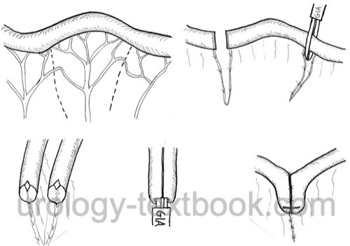
The desired intestinal loop is held against the surgical light to see the vasculature shining through the mesentery. Incise the peritoneum along the desired line on both sides [see figure bowel anastomosis with GIA]. Vessels crossing the peritoneal incision are isolated and ligated. The isolation of the bowel wall from the mesentery should be done very carefully (1 cm length) to ensure adequate blood supply of the bowel ends.
Small bowel anastomosis with GIA:
A GIA is a linear cutting and suturing stapler device. In the first step, the isolated bowel loop is resected using two applications (two magazines) of the GIA stapler. The use of GIA for bowel resection minimizes the fecal spillage of the abdominal cavity. The two bowel ends are juxtaposed in a side-to-side fashion. Resect the antimesenteric corner of each staple line (small enterostomy) so that the forks of the GIA suturing device can be inserted into each lumen of the bowel ends. Assemble the GIA device and use the third magazine to create a large side-to-side enterostomy. Avoid cutting the bowel wall near the mesentery. Remove the GIA device and close the remaining opening with a hand-sewn technique or using a fourth magazine of the stapler [see figure bowel anastomosis with GIA].
Hand-sewn bowel anastomosis
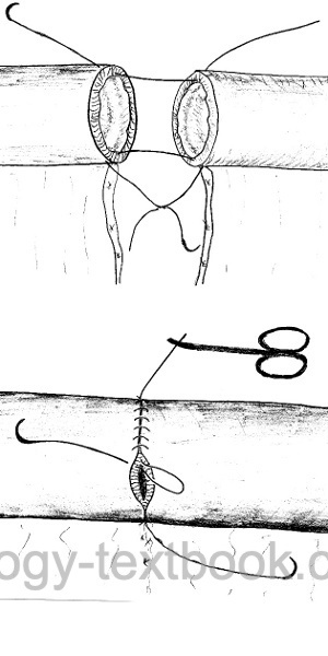
The suture material for small bowel anastomosis is Vicryl or PDS (4-0). At first, the two ends of the bowel are brought close together, and corner sutures are placed at the mesenteric and antimesenteric ends of the open bowel. Use the corner sutures for the anastomosis with a running seromuscular suturing: all intestinal wall layers except the lamina epithelialis mucosae are grasped [fig. hand-sewn bowel anastomosis]. After the half circumference, turn the anastomosis with the corner sutures and close the remaining defect with a running suture using the second corner suture. Alternatively, seromuscular bowel anastomosis can be done using an interrupted suturing technique (Kremer et al., 1992).
 |
Postoperative Care
General Measures:
- Early mobilization
- Intensive respiratory therapy
- Thrombosis prophylaxis
- Laboratory tests
- Wound checks
Analgesia:
Provide analgesia with a combination of NSAIDs and opioids. An epidural anesthesia facilitates postoperative pain management and minimizes the need for opioids.
Oral Diet after Small Bowel Anastomosis
- Remove the nasogastric tube after surgery
- Allow small sips of clear liquids after surgery
- Increase amounts of clear liquids and allow yogurt or pudding on postoperative days 1 and 2
- If the patient feels well, allow small amounts of solid food (appetite-driven) starting postoperative day 3
Complications of Bowel Anastomosis
- Intraabdominal abscess, wound infection
- Anastomotic insufficiency with peritonitis
- Postoperative paralytic ileus
- Anastomotic stenosis and mechanical ileus
- Small bowel fistula or intraabdominal abscess
| Laser treatment | Index | Simple nephrectomy |
Index: 1–9 A B C D E F G H I J K L M N O P Q R S T U V W X Y Z
References
Kremer, K.; Lierse, W.; Platzer, W.; Schreiber, H. W. & Weller, S. (ed.) Chirurgische Operationslehre: Spezielle Anatomie, Indikationen, Technik, KomplikationenThieme, 1992, Band 6 Darm.
 Deutsche Version: Technik der Dünndarm Anastomose
Urology-Textbook.com – Choose the Ad-Free, Professional Resource
This website is designed for physicians and medical professionals. It presents diseases of the genital organs through detailed text and images. Some content may not be suitable for children or sensitive readers. Many illustrations are available exclusively to Steady members. Are you a physician and interested in supporting this project? Join Steady to unlock full access to all images and enjoy an ad-free experience. Try it free for 7 days—no obligation.
New release: The first edition of the Urology Textbook as an e-book—ideal for offline reading and quick reference. With over 1300 pages and hundreds of illustrations, it’s the perfect companion for residents and medical students. After your 7-day trial has ended, you will receive a download link for your exclusive e-book.