You are here: Urology Textbook > Prostate > Prostatic cysts
Prostate cysts: Symptoms, Diagnosis and Treatment
Definition
Prostate cysts are fluid-filled cavities in the prostate, usually lined by epithelium (true cysts).
 |
Epidemiology:
5–8% of men have small prostatic cysts (Ishikawa et al., 2003).
Etiology and Pathology of Prostate Cysts
Depending on the localization, cysts are divided into median (midline cysts) or peripheral (lateral) cysts:
Utricle cyst:
A utricle cyst (or Müllerian duct cyst) presents with a midline cyst of the prostate and originates from the utricle. Histologically, a high PSA expression is typical. Squamous epithelium is not typical for a utricle cyst.
Persisting Müllerian Duct Syndrome:
Persisting Müllerian duct syndrome may cause large solid and cystic pelvic masses near the midline. Pathologic examination shows squamous epithelium analogous to the vagina and, in some cases, uterine tissue.
Other Causes of Prostatic Cysts:
Cysts derived from seminal vesicles, seminal tract or prostate gland, caused by infection, BPH or malignant tumors (Galosi et al., 2009).
Signs and Symptoms of Prostatic Cysts
Small cystic lesions are usually asymptomatic and clinically insignificant. Larger cysts may cause:
- Lower urinary tract symptoms due to subvesical obstruction
- Urinary retention
- Hydronephrosis
- Urinary tract infection
- Hematospermia
Diagnostic Workup
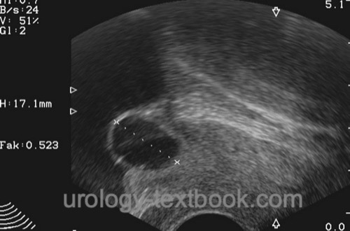
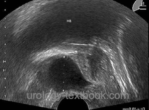
- Transrectal ultrasonography [fig. prostatic cyst in TRUS]
- Abdominal ultrasonography: pelvic masses? Residual urine?
- Urinary flow test
- Cystoscopy
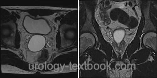
- CT or MRI for larger or complex cysts (Curran et al., 2007)
 |
 |
 |
Differential Diagnosis of Prostatic Cysts
Defect after TURP, diverticulum or bladder or urethra, prominent seminal vesicles, Ectopic ureter or ectopic ureterocele.
Treatment of Prostatic Cysts
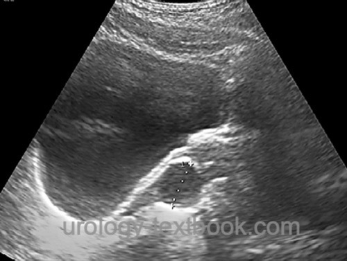
Small symptomatic prostatic cysts can be treated by transurethral resection [fig. transurethral deroofing of a prostatic cyst]. Large pelvic masses of the Müllerian duct system may need open surgery or laparoscopic surgery. Typical open surgical approaches are either a lower abdominal incision with a transvesical-transtrigonal approach or the perineal approach.
| Prostate diseases | Index | Prostatitis |
Index: 1–9 A B C D E F G H I J K L M N O P Q R S T U V W X Y Z
References
S. Curran, O. Akin, A. M. Agildere, J. Zhang, H. Hricak, and J. Rademaker, “Endorectal MRI of prostatic and periprostatic cystic lesions and their mimics.,” AJR. American journal of roentgenology, vol. 188, no. 5, pp. 1373–1379, 2007.
Galosi, A. B.; Montironi, R.; Fabiani, A.; Lacetera, V.; Gallé, G. & Muzzonigro, G.
Cystic lesions of the prostate gland: an ultrasound classification with pathological correlation.
J Urol, 2009, 181, 647-657
M. Ishikawa et al., “Midline Prostatic Cysts in Healthy Men: Incidence and Transabdominal Sonographic Findings,” American Journal of Roentgenology, vol. 181, no. 6, pp. 1669–1672, 2003.
M. Ishikawa et al., “Midline prostatic cysts in healthy men: incidence and transabdominal sonographic findings.,” AJR. American journal of roentgenology, vol. 181, no. 6, pp. 1669–1672, 2003.
 Deutsche Version: Ursachen und Therapie von Prostatazysten
Urology-Textbook.com – Choose the Ad-Free, Professional Resource
This website is designed for physicians and medical professionals. It presents diseases of the genital organs through detailed text and images. Some content may not be suitable for children or sensitive readers. Many illustrations are available exclusively to Steady members. Are you a physician and interested in supporting this project? Join Steady to unlock full access to all images and enjoy an ad-free experience. Try it free for 7 days—no obligation.
New release: The first edition of the Urology Textbook as an e-book—ideal for offline reading and quick reference. With over 1300 pages and hundreds of illustrations, it’s the perfect companion for residents and medical students. After your 7-day trial has ended, you will receive a download link for your exclusive e-book.