You are here: Urology Textbook > Anatomy > Penis
Penis: Anatomy, Vascular Supply, and Histology
- Anatomy of the Penis: gross appearance, vascular supply, histology.
- Mechanisms of penile erection: phases of erection, innervation, neural control, molecular signaling.
References: (Benninghoff, 1993 ).
Gross Anatomy of the Penis
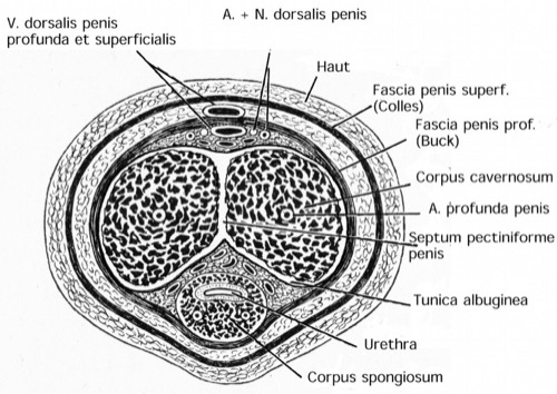 |
Transverse section of the penis. Figure modified from Gray's Anatomy, Lea and Febiger 1918, Philadelphia, USA. |
Corpora of the Penis
The penis is composed of two corpora cavernosa and one unpaired corpus spongiosum. The corpora contain oddly shaped cavities, which are lined with endothelium. Smooth muscle runs through the walls and septa of the corpora. The afferent arteries are notable for their strong smooth muscle wall.
Corpora cavernosa penis:
Each corpora cavernosa begins at the inferior ramus of the pubic bone (crura penis) and extends through the penis to the glans. The corpora cavernosa are surrounded by the ischiocavernosus muscle.
Corpus spongiosum penis:
The unpaired corpus spongiosum surrounds the urethra and begins between the two crura penis as a thickening (Bulbus penis). At the tip of the penis, the corpus spongiosum forms the glans penis. Connective tissue fibers provide a strong connection to the corpora cavernosa. The M. bulbocavernosus surrounds the bulb of the penis.
Glans penis:
The glans penis is the conical shaped end of the corpus spongiosum. The urethra ends with the external urethral orifice or meatus urethrae externus at the tip of the glans. The Corona glandis forms a sulcus and marks the transition from the corpus spongiosum to the corpus cavernosum.
Tunica albuginea of the penis:
The tunica albuginea is a strong 1–2 mm thick fascial sheath; it surrounds the outside of the two corpora cavernosa and forms a septum in between (Septum pectiniforme penis).
Fascia and Ligaments of the Penis
The corpora of the penis are circularly surrounded by the Buck fascia (Fascia penis profunda). The fascia penis superficialis lies above the Buck fascia and is endorsed with smooth muscle from the tunica dartos [Figure: transverse section of the penis]. The fascia penis superficialis connects caudal to the superficial perineal fascia (Colles' fascia) and cranial to the Scarpa's fascia. The connections of the fascia penis superficialis restrict penile hematomas to the area between the inguinal ligament, the fascia lata, and the ventral of the central tendon of the pelvic floor.
The penile ligaments attach the penis to the symphysis and the linea alba of the rectus sheath (fundiform ligament and suspensory ligament).
Prepuce (Foreskin)
The prepuce (foreskin) consists of an inner and outer leaf and covers the glans of the flaccid penis. The foreskin serves as reserve skin during erection. The two skin sheets are movable against each other. The frenulum is located at the ventral side of the glans and attaches the prepuce to the glans [fig. uncircumcised penis]. The smegma is whitish pollution of the glans and prepuce and arises due to bacterial colonization of the desquamated epithelium.
Anatomy of the Male Urethra
The male urethra extends from the bladder neck (meatus urethrae internus) to the meatus (urethrae externus) and can be divided into the following sections:
Prostatic urethra:
Surrounded by the prostate, 3–5 cm long. The colliculus seminalis is located in the middle of the prostatic urethra. At the colliculus seminalis, also called verumontanum, end the ejaculatory ducts and pass into the urethra. The prostatic utricle is a relic of the embryonic period (Müllerian duct) in the midline of the prostatic urethra.
Membranous urethra:
The membranous urethra runs through the pelvic floor and is enclosed by the external urethral sphincter (M. sphincter urethrae externum).
Spongy urethra:
Consists of the bulbar and penile urethra, the distal end of the ischiocavernosus and bulbospongiosus muscles define the transition. The Cowper glands drain into the proximal spongy urethra. The urethral glands (Littre's glands) open on the entire length of the urethra.
Fossa navicularis:
The fossa navicularis is an increase in diameter of the penile urethra while transversing the glans.
Vascular Supply of the Penis
Arteries of the penis:
All penile arteries arise from the internal pudendal artery from the internal iliac artery [Fig. Arteries of the penis]. The internal pudendal artery reaches the ischiorectal fossa via Alcock's canal.
Dorsal artery of penis:
The dorsal artery of the penis is located on the dorsum of the corpus cavernosum and supplies the glans, the prepuce and penile skin. Latin: A. dorsalis penis.
Deep artery of penis:
The deep artery of penis is located in the center of the corpus cavernosum and emits many Aa. helicinae (helicine arteries) which flow into the caverns of the corpus cavernosum. Latin: A. profunda penis.
Bulbourethral artery:
The bulbourethral artery (latin: A. bulbi penis) supplies the corpus spongiosum and the urethra.
Veins of the penis:
The veins of the penis break through the tunica albuginea and open via the circumflex veins in the unpaired deep dorsal vein, which empties into the venous plexus of Santorini. The superficial dorsal vein lies external to Buck fascia.
Penile Veins:
The veins of the penis pass through the tunica albuginea and drain via the circumflex veins into the unpaired deep dorsal vein of the penis. The deep veins below the Buck fascia empty into the prostatic venous plexus (Santorini plexus) and then into the pelvic venous system. The superficial dorsal vein of the penis drains into the external pudendal veins.
Lymphatic vessels of the penis:
Drain into the superficial inguinal and subinguinal lymph nodes. The proximal urethra drains into the pelvic lymph nodes (internal and common iliac lymph nodes).
Innervation of the Penis
See next section mechanisms of penile erection.
Histology of the Penis
Histology of the male urethra:
The layers of the urethra consists of:
- mucosa with varying epitheliums (prostatic urethra with transitional epithelium, spongy urethra with stratified columnar epithelium, fossa navicularis with squamous epithelium),
- submucosa with connective tissue and glands,
- and a very thin muscularis.
Histology of the Glans and prepuce:
Keratinized squamous epithelium.
Histology of the erectile tissue:
The corpora contain septa of smooth muscle, which form bizarrely shaped cavities. Vascular endothelium lines the septa and cavities. The afferent arteries for the erectile tissue possess a strong muscularis.
| Anatomy | Index | Penile Erection |
Index: 1–9 A B C D E F G H I J K L M N O P Q R S T U V W X Y Z
References
Benninghoff, A. (1993): Makroskopische Anatomie, Embryologie und Histologie des Menschen. 15. Auflage. München; Wien; Baltimore: Urban & Schwarzenberg.
Porst 2004 PORST, H.:
Tadalafil, Therapiestrategien bei erektiler Dysfunktion.
Linkenheim-Hochstetten : Aesopus Verlag, 2004
 Deutsche Version: Anatomie, Arterien, Venen und Histologie des Penis.
Urology-Textbook.com – Choose the Ad-Free, Professional Resource
This website is designed for physicians and medical professionals. It presents diseases of the genital organs through detailed text and images. Some content may not be suitable for children or sensitive readers. Many illustrations are available exclusively to Steady members. Are you a physician and interested in supporting this project? Join Steady to unlock full access to all images and enjoy an ad-free experience. Try it free for 7 days—no obligation.
New release: The first edition of the Urology Textbook as an e-book—ideal for offline reading and quick reference. With over 1300 pages and hundreds of illustrations, it’s the perfect companion for residents and medical students. After your 7-day trial has ended, you will receive a download link for your exclusive e-book.