You are here: Urology Textbook > Bladder > Bladder injury
Diagnosis and Treatment of Bladder Injury
Definition and Epidemiology of Bladder Injury
Injuries of the urinary bladder are caused by pelvic fracture, blunt or penetrating trauma, or iatrogenic injury (Machtens u.a., 2000). EAU Guidelines: Urological Trauma.
AAST classification of bladder trauma:
- Grade I: contusion, hematoma or partial thickness laceration of the bladder wall
- Grade II: extraperitoneal bladder wall laceration <2 cm
- Grade III: extraperitoneal bladder wall laceration >2 cm or intraperitoneal laceration <2 cm
- Grade IV: intraperitoneal bladder wall laceration >2 cm
- Grade V: bladder wall laceration extending into the bladder neck or trigone
Etiology (Causes) of Bladder Injury
Pelvic Fracture:
Injury of the bladder due to a pelvic fracture is caused by perforation of the bladder by sharp fracture fragments. The location of the bladder injury is usually extraperitoneal.
Bladder Rupture:
A filled bladder exposed to blunt trauma of the lower abdomen (most common high-velocity accidents) may rupture due to the sudden pressure increase. The location of the bladder perforation is usually intraperitoneal at the bladder dome.
Penetrating Injury:
Stab or gunshot wounds to the lower abdomen cause intraperitoneal bladder injury and concurrent injury of the bowel or large vessels.
Iatrogenic injury:
By pelvic or transurethral surgery: cesarean section, vaginal hysterectomies, retropubic slings and transurethral resection of the bladder.
Signs and Symptoms of Bladder Injury
Pelvic fracture:
Pelvic pain, pelvic instability, shock, gluteal hematoma, and abdominal tenderness.
Bladder Injury:
Hematuria, oliguria, lower abdominal pain, peritonitis, increasing retention parameters due to the resorption of urine.
Diagnostic Workup in Suspected Bladder Injury
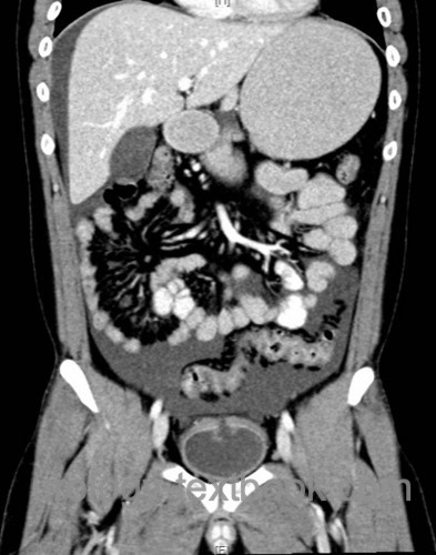
Classic imaging of suspected bladder injury includes a pelvis X-ray, retrograde urethrography, and cystography.
An abdominal CT scan with a contrast medium-filled bladder can substitute the mentioned imaging studies (except for retrograde urethrography) and provide additional information about concurrent injury.
 |
Treatment of Bladder Injury
Conservative Treatment:
Small extraperitoneal bladder injuries can be treated by inserting a transurethral catheter only. Noteworthy is to exclude bone fragments that protrude into the bladder and continue to maintain bladder perforation.
Surgical Treatment:
Surgical treatment is necessary for large extraperitoneal bladder injuries and all intraperitoneal bladder ruptures. A midline laparotomy to the lower abdomen is done. Check the abdominal cavity for further injury. The bladder dome is opened, and the bladder is inspected for perforation. The injury is closed in three layers (mucosa – muscularis – peritoneum).
| Bladder exstrophy | Index | Urethral caruncle |
Index: 1–9 A B C D E F G H I J K L M N O P Q R S T U V W X Y Z
References
EAU Guidelines: Urological Trauma.
Machtens u.a. 2000 MACHTENS, S.. ; STIEF,
C. G. ; HAGEMANN, J. ; PFINGST, G. ; GäNSSLEN,
A. ; POHLEMANN, T. ; TRUSS, M. C. ; KUCZYK,
M. A. ; BECKER, A. J. ; JONAS, U.:
Management traumatischer Läsionen von Harnblase und Urethra.
In: Urologe B
40 (2000), S. 560–571
 Deutsche Version: Verletzung der Harnblase
Urology-Textbook.com – Choose the Ad-Free, Professional Resource
This website is designed for physicians and medical professionals. It presents diseases of the genital organs through detailed text and images. Some content may not be suitable for children or sensitive readers. Many illustrations are available exclusively to Steady members. Are you a physician and interested in supporting this project? Join Steady to unlock full access to all images and enjoy an ad-free experience. Try it free for 7 days—no obligation.
New release: The first edition of the Urology Textbook as an e-book—ideal for offline reading and quick reference. With over 1300 pages and hundreds of illustrations, it’s the perfect companion for residents and medical students. After your 7-day trial has ended, you will receive a download link for your exclusive e-book.