You are here: Urology Textbook > Anatomy > Testes
Testis, Epididymis and Spermatogenesis: Histology
- Anatomy of the Testes: Gross appearance, vascular supply, innvervation, spermatic cord.
- Anatomy of the Testes: Histology, Spermatogenesis.
- Anatomy of the Testes: Male Hormones (Testosteron, LH, FSH, Inhibin).
References: (Benninghoff, 1993).
Histology of the testis
The seminiferous tubules have a diameter of about 200 μm and contain the germinal epithelium. One or several highly convoluted seminiferous tubules form a lobule of the testis. The spermatogonia (single: spermatogonium) form the basal layer of the germinal epithelium and are a kind of the stem cells of the spermatocytes production. Per hour, approximately one million sperm cells are produced and passed to the Epididymis. The time period from of a spermatogonium to the spermatocyte in the semen takes approximately 60–80 days.
Meiosis:
spermatocytes I arise by mitotic divisions from spermatogonia and contain the normal diploid set of chromosomes (46,XY). The first meiotic division (prophase, metaphase, anaphase and telophase I) of spermatocytes I leads to two spermatocytes II (23,X or Y). The Prophase I is divided into the following sub-steps:
Leptotene:
The DNA is condensed.
Zygotene:
Homologous chromosomes line up (conjugation).
Pachytene:
Coiling of the paired chromosomes and exchange of genetic material (crossing over).
Diplotene:
Crossing over is visible in the light microscope.
Diakinesis:
Resolution of the nuclear membrane and the initiation of metaphase I. After completion of the first meiotic division of the spermatocyte II has 22 autosomes and one sex chromosome, X or Y. Each chromosome has two chromatids (2n).
The second meiotic division (prophase, metaphase, anaphase and telophase II) divides Spermatocytes II into two spermatids, without doubling the DNA content. A spermatid therefore has 22 autosomes and one sex chromosome, each haploid chromosome consists of only one DNA strand (23,X or Y, 1n).
Development of spermatozoa:
A complex differentiation leads from the first round spermatid to the mature spermatozoa. Following steps are distinguished:
Nuclear condensation to one-tenth of the initial volume
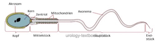
Acrosome formation: cap-shaped structure at the head of the sperm containing the enzymes hyaluronidase and acrosin. The acrosome allows the penetration of the sperm into the ovum through the zona pellucida.
Tail system: eukaryontic flagellum based on two single central and nine peripheral microtubule doublets (Axoneme). In the mid piece there are plenty mitochondria, the main piece of the flagellum is surrounded by a ring-shaped fibers.
Following the delivery of spermatozoa to the epididymis, the spermatozoa are still immobile and cannot fertilize an ovum. Only after the maturation process in the epididymis (spermiogenesis), they become mobile and capable of fertilization.
Anatomy of the spermatozoa:
The mature sperm cells consist of head and tail. The head has a size of approximately 2 μm wide and 4 μm in length; it contains the nucleus and is covered by the acrosome. The tail has a length of about 60 μm; it is divided into a mid piece with lots of mitochondria, and a tail [fig. spermatozoen]. See section semen analysis for reference values of spermatozoa.
 |
Sertoli cells:
During embryogenesis, the Sertoli cells secrete the antimullerian hormone, which prevents the development of the female reproductive organs. The fully developed Sertoli cells surround the germ cells and have important tasks in support and nutrition of germ cells, formation of the blood-testis barrier, phagocytosis of spermatid remains, secretion of inhibin (control of FSH), and transmission of hormonal stimuli.
Sertoli cells are located on the basement membrane of seminiferous tubules and reach with its cytoplasm to luminal. Sertoli cells embed with their cytoplasm the cells of the spermatogenesis. The nucleus is near the basement membrane. Sertoli cells cannot divide, the number remains constant even after damage to the germinal epithelium.
Leydig cells:
Leydig cells are found between the seminiferous tubules in the testes. They produce testosterone in response to LH. Testosterone is produced in the smooth endoplasmic reticulum of the leydig cell.
Rete testis:
Anastomosing network of tubules, lined with single-layer isoprismatic epithelium. The seminiferous tubules start and end at the rete testis. The germinal epithelium suddenly ends with begin of the rete testis, forming a kind of valve to prevent the backflow of spermatozoa.
Histology of the epididymis and vas deferens
The efferent ductules of the epididymis are lined by a columnar epithelium with cilia and microvilli. The epithelium absorbs the testicular fluid and provides further transport of spermatozoa. The ductus epididymidis is lined by columnar epithelium with stereocilia and has a wall of smooth muscle cells. The passage of spermatozoa through the epididymis takes 8–17 days, during this period a maturation process takes place (see above).
Ductus deferens (vas deferens) is lined by a pseudostratified prismatic epithelium with stereocilia. The smooth muscle wall is strong and consists of three layers (longitudinal-transverse-longitudinal).
| Anatomy Testis | Index | Male sex hormones |
Index: 1–9 A B C D E F G H I J K L M N O P Q R S T U V W X Y Z
References
Benninghoff 1993 BENNINGHOFF, A.: Makroskopische Anatomie, Embryologie und Histologie des Menschen.15. Auflage.
München; Wien; Baltimore : Urban und Schwarzenberg, 1993
 Deutsche Version: Histologie der Hoden und der Spermatogenese.
Urology-Textbook.com – Choose the Ad-Free, Professional Resource
This website is designed for physicians and medical professionals. It presents diseases of the genital organs through detailed text and images. Some content may not be suitable for children or sensitive readers. Many illustrations are available exclusively to Steady members. Are you a physician and interested in supporting this project? Join Steady to unlock full access to all images and enjoy an ad-free experience. Try it free for 7 days—no obligation.
New release: The first edition of the Urology Textbook as an e-book—ideal for offline reading and quick reference. With over 1300 pages and hundreds of illustrations, it’s the perfect companion for residents and medical students. After your 7-day trial has ended, you will receive a download link for your exclusive e-book.